WB - 1:1000, IHC - 1:100 to 1:500. Epitope retrieval with citrate buffer pH6.0 is recommended for FFPE tissue sections. Zinc-fixative (JB fix) is recommended to enhance staining., ICC - 1:100 to 1:500. Epitope retrieval with citrate buffer pH6.0 is recomm
Application Notes:
Application Notes: Format: Whole IgG
Detection of mouse MafA by immunohistochemistry. Sample: FFPE section of mouseBeta-TC-6 cells. Antibody: Rabbit anti-MafA recombinant monoclonal antibody (orb1519835). Secondary: HRP-conjugated goat anti-rabbit IgG.
Detection of mouse MafA by immunohistochemistry. Sample: FFPE section of mouse pancreas (JB fixation). Antibody: Rabbit anti-MafA recombinant monoclonal antibody (orb1519835). Secondary: HRP-conjugated goat anti-rabbit IgG.
Detection of human MafA by immunohistochemistry. Sample: FFPE section of human pancreatic tumor. Antibody: Rabbit anti-MafA recombinant monoclonal antibody (orb1519835). Secondary: HRP-conjugated goat anti-rabbit IgG.
Detection of human MafA (red) by immunohistochemistry. Sample: FFPE section of human pancreatic carcinoma. Antibody: Rabbit anti-MafA recombinant monoclonal antibody (orb1519835) used at 1:250. Secondary: HRP-conjugated goat anti-rabbit IgG. Substrate: Opal(TM). Counterstain: DAPI (blue).
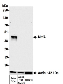
Detection of mouse MafA by western blot. Samples: Whole cell lysate (10 µg) from Beta-TC-6, AlphaTC1 Clone 9, and NIH 3T3 cells prepared using NETN lysis buffer. Antibody: Rabbit anti-MafA recombinant monoclonal antibody (orb1519835) used at 1:1000. Secondary: HRP-conjugated goat anti-rabbit IgG. Detection: Chemiluminescence with an exposure time of 10 seconds. Lower Panel: Rabbit anti-Actin recombinant monoclonal antibody.
Detection of human MafA by western blot. Samples: Whole cell lysate (10 µg) from HEK293T transfected with myc tagged Human MafA, Human MafB, and Human cMaf prepared using NETN lysis buffer. Antibody: Rabbit anti-MafA recombinant monoclonal antibody (orb1519835) used at 1:1000. Secondary: HRP-conjugated goat anti-rabbit IgG. Detection: Chemiluminescence with an exposure time of 30 seconds. Lower Panel: Rabbit recombinant monoclonal antibody to Myc-tag.
Detection of mouse MafA by western blot. Samples: Whole cell lysate (10 µg) from HEK293T transfected with myc tagged mouse MafA, Human MafB, and Human cMaf prepared using NETN lysis buffer. Antibody: Rabbit anti-MafA recombinant monoclonal antibody (orb1519835) used at 1:1000. Secondary: HRP-conjugated goat anti-rabbit IgG. Detection: Chemiluminescence with an exposure time of 30 seconds. Lower Panel: Rabbit recombinant monoclonal antibody to Myc-tag.
* VAT and and shipping costs not included. Errors and price changes excepted