Borate Buffered Saline (BBS) pH 8.2 with 0.1% rAlbumin and 0.09% Sodium Azide
Purity:
Recombinant antibody was purified from cell culture supernatant
Form:
Liquid
Target:
LAG3
Application Dilute:
Flow-Cyt Fixed in 4% formaldehyde and permeabilized with 90% methanol. 1 µl per 1 x 10 6 cells. Immunocytochemistry (ICC) 1:100 to 1:500. Epitope retrieval with citrate buffer pH6.0 is recommended for FFPE cell sections. Immunohistochemistry (IHC) 1:100 t
Application Notes:
Application Notes: All western blot analysis is performed using 5% Milk-TBST for blocking and as antibody diluent. Primary antibody is incubated overnight
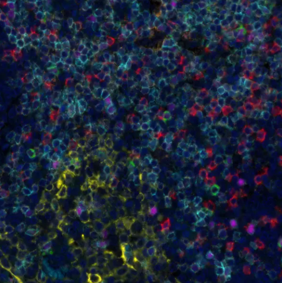
Detection of human LAG3 (red) by immunohistochemistry. Sample: FFPE section of human tonsil. Antibody: Rabbit anti-LAG3 recombinant monoclonal antibody (orb1519933) used at 1:250. Secondary: HRP-conjugated goat anti-rabbit IgG. Substrate: Opal(TM). Counterstain: DAPI (blue).
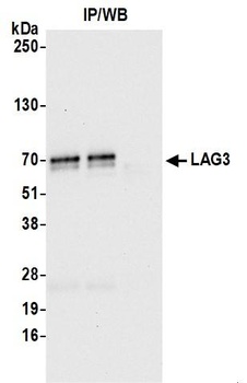
Detection of human LAG3 by western blot of immunoprecipitates. Samples: Whole cell lysate (1.0 mg per IP reaction, 20% of IP loaded) from HDLM-2 cells prepared using NETN lysis buffer. Antibodies: Rabbit anti-LAG3 recombinant monoclonal antibody (orb1519933) used for IP at 12 µl per reaction.
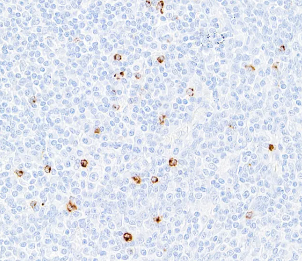
Detection of human LAG3 in FFPE tonsil by immunohistochemistry. Antibody: Rabbit anti-LAG3 recombinant monoclonal antibody. Secondary: HRP-conjugated goat anti-rabbit IgG. Substrate: DAB.
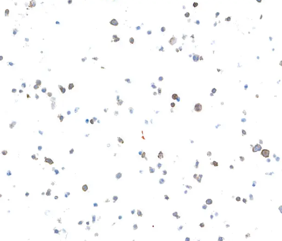
Detection of human LAG3 in FFPE HDLM-2 by immunocytochemistry. Antibody: Rabbit anti-LAG3 recombinant monoclonal antibody. Secondary: HRP-conjugated goat anti-rabbit IgG. Substrate: DAB
* VAT and and shipping costs not included. Errors and price changes excepted