Borate Buffered Saline (BBS) pH 8.2 with 0.1% rAlbumin and 0.09% Sodium Azide
Form:
Liquid
Target:
KAP-1 Phospho (S824)
Application Dilute:
WB - 1:1000, IP - 20 µl/mg lysate, IHC - 1:100 - 1:500. Epitope retrieval with citrate buffer pH 6.0 is recommended for FFPE tissue sections., ICC-IF - 1:100 - 1:500. Formaldehyde fixation is recommended. Permeabilization with Triton-X 100 is recommended
Application Notes:
Application Notes: Format: Whole IgG
Detection of human phospho KAP-1 (shaded) in etoposide treated HEK293T cells (right) and untreated HEK293T cells (left) by flow cytometry. Antibody: Rabbit anti-phospho KAP-1 recombinant monoclonal [BL-246-7B5] (orb1519972) or isotype control (unshaded). Secondary: DyLight 488-conjugated goat anti-rabbit IgG.
Detection of human Phospho KAP-1 (S824) in FFPE etoposide treated HeLa cells by ICC. Mock phosphatase treated section (left) and calf intestinal phosphatase-treated section (right). Antibody: Rabbit anti-Phospho KAP-1 (S824) recombinant monoclonal [BL-246-7B5] (orb1519972). Secondary: HRP-conjugated goat anti-rabbit IgG. Substrate: DAB.
Detection of human Phospho KAP-1 (S824) by immunocytochemistry. Samples: Formaldehyde-fixed asynchronous HeLa cells grown in chambered microscope slides and treated with etoposide (right) or untreated (left). Antibody: Rabbit anti-Phospho KAP-1 (S824) recombinant monoclonal antibody [BL-246-7B5] (orb1519972) used at of 1:100. Secondary: DyLight 594-conjugated goat anti-rabbit IgG. Counterstain: Phalloidin conjugated Alexa Fluor 488 (green).
Detection of human Phospho KAP-1 (S824) in FFPE prostate carcinoma by IHC. Mock phosphatase treated section (left) and calf intestinal phosphatase-treated section (right). Antibody: Rabbit anti-Phospho KAP-1 (S824) recombinant monoclonal [BL-246-7B5] (orb1519972). Secondary: HRP-conjugated goat anti-rabbit IgG. Substrate: DAB.
Detection of human Phospho KAP-1 (S824) by western blot of immunoprecipitates. Samples: Whole cell lysate (1.0 mg per IP reaction, 5% of IP loaded) from HEK293T cells prepared using NETN lysis buffer that were treated with 100 µM etoposide (+) or mock treated (-). Antibodies: Rabbit anti-Phospho KAP-1 (S824) recombinant monoclonal antibody [BL-246-7B5] (orb1519972) used for IP at 20 µl/mg lysate.
Detection of human Phospho KAP-1 (S824) by Simple Western(TM). Samples: Whole cell lysate (0.4 mg/mL) from HEK293T cells treated with 100 µM etoposide prepared using NETN lysis buffer. Antibody: Rabbit anti-Phospho KAP-1 (S824) recombinant monoclonal antibody [BL-246-7B5] (orb1519972) used at 1:10, 1:50, and 1:250. Separation and Detection: SallySue ProteinSimple instrument with the 12-230 kDa separation module and anti-Rabbit detection module. Left Panel: Virtual Lane View. Right Panel: Electropherogram.
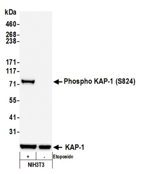
Detection of mouse Phospho KAP-1 (S824) by western blot. Samples: Whole cell lysate (50 µg) from NIH3T3 cells treated with 100 µM etoposide (+) or mock treated (-) prepared using NETN lysis buffer. Antibody: Rabbit anti-Phospho KAP-1 (S824) recombinant monoclonal antibody [BL-246-7B5] (orb1519972) used at 1:1000. Secondary: HRP-conjugated goat anti-rabbit IgG. Chemiluminescence with an exposure time of 3 seconds. Lower Panel: Rabbit anti-KAP1 recombinant monoclonal antibody [BL-248-2G6].
Detection of human Phospho KAP-1 (S824) by western blot. Samples: Whole cell lysate (25 µg) from HEK293T cells treated with 100 µM etoposide (+) or mock treated (-) prepared using NETN lysis buffer. Antibody: Rabbit anti-Phospho KAP-1 (S824) recombinant monoclonal antibody [BL-246-7B5] (orb1519972) used at 1:1000. Secondary: HRP-conjugated goat anti-rabbit IgG. Chemiluminescence with an exposure time of 3 seconds. Lower Panel: Rabbit anti-KAP1 recombinant monoclonal antibody [BL-248-2G6].
* VAT and and shipping costs not included. Errors and price changes excepted