cell cycle and apoptosis regulator protein 2, cell division cycle and apoptosis regulator protein 2, DBC.1, DBC1, DBC-1, DBIRD complex subunit KIAA1967, deleted in breast cancer 1, Deleted in breast cancer gene 1 protein, KIAA1967, NET35, p30 DBC, p30 DBC protein, p30DBC
Tris-citrate/phosphate buffer, pH 7 to 8 containing 0.09% Sodium Azide
Form:
Liquid
Target:
DBC1/p30 DBC
Application Dilute:
WB - 1:2,000 - 1:10,000, IP - 2 - 10 µg/mg lysate, IHC - 1:1,000 - 1:5,000. Epitope retrieval with citrate buffer pH 6.0 is recommended for FFPE tissue sections.
Application Notes:
Application Notes: Format: Whole IgG
Detection of human DBC1/p30 by immunohistochemistry.Sample: FFPE section of human prostate carcinoma. Antibody: Affinity purified rabbit anti-BC1/p30 (Cat. No. orb1523889) used at a dilution of 1: 1, 000 (1µg/ml). Detection: DAB
Detection of mouse DBC1/p30 by immunohistochemistry.Sample: FFPE section of mouse teratoma. Antibody: Affinity purified rabbit anti-BC1/p30 (Cat. No. orb1523889) used at a dilution of 1: 1, 000 (1µg/ml). Detection: DAB
Detection of human DBC1/p30 by western blot of immunoprecipitates. Samples: Whole cell lysate (1 mg for IP, 20% of IP loaded) from HEK293T cells. Antibodies: Affinity purified goat anti-DBC1/p30 antibody orb1523889 used for IP at 6 µg/mg lysate.
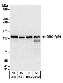
Detection of human DBC1/p30 by western blot. Samples: Whole cell lysate from HEK293T (15 and 50 µg) , HeLa (50µg) , and Jurkat (50µg) cells. Antibodies: Affinity purified goat anti-DBC1/p30 antibody orb1523889 used for WB at 0.1 µg/ml. Detection: Chemiluminescence with an exposure time of 30 seconds.
Detection of mouse DBC1/p30 by western blot. Samples: Whole cell lysate (50 µg) from NIH 3T3, Renca, and TCMK-1 cells. Antibodies: Affinity purified goat anti-DBC1/p30 antibody orb1523889 used for WB at 0.1 µg/ml. Detection: Chemiluminescence with an exposure time of 30 seconds.
* VAT and and shipping costs not included. Errors and price changes excepted