
5HT1F Receptor Rabbit Polyclonal Antibody, Unconjugated
Catalog Number:
BYT-ORB155530
- Images (8)


| Article Name: | 5HT1F Receptor Rabbit Polyclonal Antibody, Unconjugated |
| Biozol Catalog Number: | BYT-ORB155530 |
| Supplier Catalog Number: | orb155530 |
| Alternative Catalog Number: | BYT-ORB155530-50,BYT-ORB155530-100,BYT-ORB155530-200 |
| Manufacturer: | Biorbyt |
| Host: | Rabbit |
| Category: | Antikörper |
| Application: | IF, IHC-Fr, IHC-P, WB |
| Species Reactivity: | Human, Mouse, Rat |
| Immunogen: | KLH conjugated synthetic peptide derived from human 5HT1F Receptor/SR-1F (1-100/366aa) |
| Conjugation: | Unconjugated |
| Alternative Names: | 5-HT-1F, 5-HT1F, 5HT6, HTR1EL, MR77, Htr1eb, 5HT1F_HUMAN, HTR1F, Serotonin receptor 1F, 5HT1F_MOUSE, 5-HT-1E-beta, 5ht1f, 5HT1F_RAT, |
| 5HT1F Receptor Rabbit Polyclonal Antibody |
| Clonality: | Polyclonal |
| Concentration: | 1mg/ml |
| Molecular Weight: | 42 kDa |
| UniProt: | P30939 |
| Buffer: | 0.01M TBS (pH7.4) with 1% rAlbumin, 0.02% Proclin300 and 50% Glycerol. |
| Form: | Liquid |
| Target: | HTR1F |
| Application Dilute: | WB=1:500-2000, IHC-P=1:100-500, IHC-F=1:100-500, IF=1:100-500 |
|
|
Immunohistochemical staining of human meningioma tissue using 5HT1F Receptor antibody. |
|
|
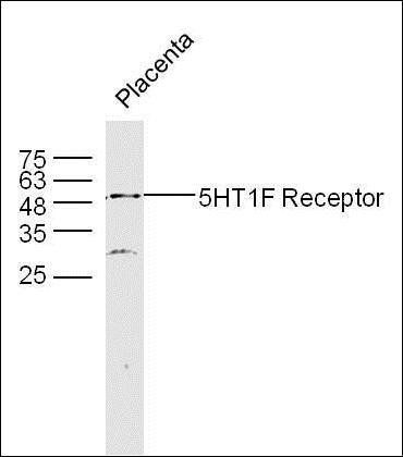
Western blot analysis of placenta lyates using 5HT1F Receptor antibody. |
|
|
Paraformaldehyde-fixed, paraffin embedded (Mouse brain), Antigen retrieval by boiling in sodium citrate buffer (pH6.0) for 15 min, Block endogenous peroxidase by 3% hydrogen peroxide for 20 minutes, Blocking buffer (normal goat serum) at 37C for 30 min, Antibody incubation with (5HT1F Receptor) Polyclonal Antibody, Unconjugated (orb155530) at 1:400 overnight at 4C, followed by operating according to SP Kit (Rabbit) instructionsand DAB staining. |
|
|
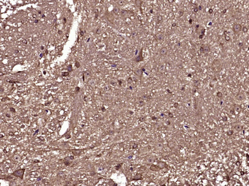
Paraformaldehyde-fixed, paraffin embedded (Mouse spinal cord), Antigen retrieval by boiling in sodium citrate buffer (pH6.0) for 15 min, Block endogenous peroxidase by 3% hydrogen peroxide for 20 minutes, Blocking buffer (normal goat serum) at 37C for 30 min, Antibody incubation with (5HT1F Receptor) Polyclonal Antibody, Unconjugated (orb155530) at 1:400 overnight at 4C, followed by operating according to SP Kit (Rabbit) instructionsand DAB staining. |
|
|
Sample: A549 Cell (Human) Lysate at 40 ug, Primary: Anti-5HT1F Receptor (orb155530) at 1/300 dilution, Secondary: IRDye800CW Goat Anti-Rabbit IgG at 1/20000 dilution, Predicted band size: 42 kD, Observed band size: 42 kD. |
|
|
Sample: Lane 1: Mouse Cerebrum tissue lysates, Lane 2: Rat Cerebrum tissue lysates, Lane 3: Rat Eye tissue lysates, Primary: Anti-5HT1F Receptor (orb155530) at 1/1000 dilution, Secondary: IRDye800CW Goat Anti-Rabbit IgG at 1/20000 dilution, Predicted band size: 42 kDa, Observed band size: 48 kDa. |
|
|
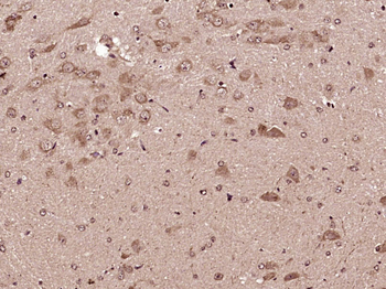
Tissue/Cell: rat brain tissue, 4% Paraformaldehyde-fixed and paraffin-embedded, Antigen retrieval: citrate buffer (0.01M, pH 6.0), Boiling bathing for 15 min, Block endogenous peroxidase by 3% Hydrogen peroxide for 30 min, Blocking buffer (normal goat serum) at 37C for 20 min, Incubation: Anti-5HT1F Polyclonal Antibody, Unconjugated (orb155530) 1:500, overnight at 4C, followed by conjugation to the secondary antibody and DAB staining. |
|
|
Tissue/Cell: rat brain tissue, 4% Paraformaldehyde-fixed and paraffin-embedded, Antigen retrieval: citrate buffer (0.01M, pH 6.0), Boiling bathing for 15 min, Blocking buffer (normal goat serum) at 37C for 20 min, Incubation: Anti-5HT1F Receptor Polyclonal Antibody, Unconjugated (orb155530) 1:200, overnight at 4C, The secondary antibody was Goat Anti-Rabbit IgG, Cy3 conjugated (orb868589) used at 1:200 dilution for 40 minutes at 37C. DAPI (5 ug/ml, blue) was used to stain the cell nuclei. |
Product Guarantee and Expert Support
