E.coli-derived human Recoverin/RCVRN recombinant protein (Position: Q33-K150).
Conjugation:
Unconjugated
Alternative Names:
Disintegrin and metalloproteinase domain-containing protein 28, ADAM 28, 3.4.24.-, Epididymal metalloproteinase-like, disintegrin-like, and cysteine-rich protein II, eMDC II, Metalloproteinase-like, disintegrin-like, and cysteine-rich protein L, MDC-L, ADAM28, ADAM23, MDCL
Anti-Recoverin/RCVRN Antibody. Tested in ELISA, IHC, WB applications. This antibody reacts with Human.
Clonality:
Polyclonal
Concentration:
Adding 0.2 ml of distilled water will yield a concentration of 500 µg/ml.
Western blot, 0.25-0.5 µg/ml, Human Immunohistochemistry(Paraffin-embedded Section), 2-5 µg/ml, Human ELISA, 0.1-0.5 µg/ml, -
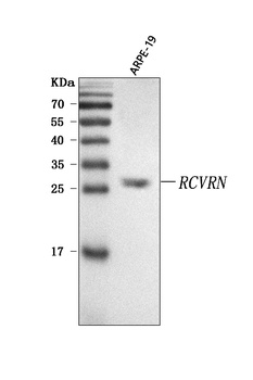
WB analysis of Recoverin/RCVRN using anti-Recoverin/RCVRN antibody.Lane 1:human ARPE-19 cell.
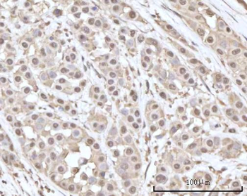
IHC analysis of Recoverin/RCVRN using anti-Recoverin/RCVRN antibody. Recoverin/RCVRN was detected in a paraffin-embedded section of human breast cancer tissue. Heat mediated antigen retrieval was performed in EDTA buffer (pH8.0, epitope retrieval solution). The tissue section was blocked with 10% goat serum. The tissue section was then incubated with 2 µg/ml rabbit anti-Recoverin/RCVRN Antibody overnight at 4C. Peroxidase Conjugated Goat Anti-rabbit IgG was used as secondary antibody and incubated for 30 minutes at 37C. The tissue section was developed using HRP Conjugated Rabbit IgG Super Vision Assay Kit with DAB as the chromogen.
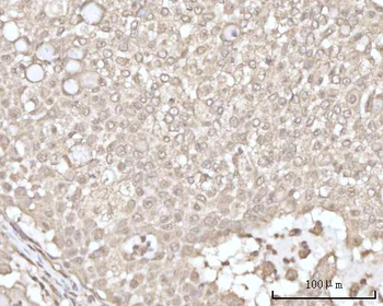
IHC analysis of Recoverin/RCVRN using anti-Recoverin/RCVRN antibody. Recoverin/RCVRN was detected in a paraffin-embedded section of human lung cancer tissue. Heat mediated antigen retrieval was performed in EDTA buffer (pH8.0, epitope retrieval solution). The tissue section was blocked with 10% goat serum. The tissue section was then incubated with 2 µg/ml rabbit anti-Recoverin/RCVRN Antibody overnight at 4C. Peroxidase Conjugated Goat Anti-rabbit IgG was used as secondary antibody and incubated for 30 minutes at 37C. The tissue section was developed using HRP Conjugated Rabbit IgG Super Vision Assay Kit with DAB as the chromogen.
IHC analysis of Recoverin/RCVRN using anti-Recoverin/RCVRN antibody. Recoverin/RCVRN was detected in a paraffin-embedded section of human ovarian cancer tissue. Heat mediated antigen retrieval was performed in EDTA buffer (pH8.0, epitope retrieval solution). The tissue section was blocked with 10% goat serum. The tissue section was then incubated with 2 µg/ml rabbit anti-Recoverin/RCVRN Antibody overnight at 4C. Peroxidase Conjugated Goat Anti-rabbit IgG was used as secondary antibody and incubated for 30 minutes at 37C. The tissue section was developed using HRP Conjugated Rabbit IgG Super Vision Assay Kit with DAB as the chromogen.
IHC analysis of Recoverin/RCVRN using anti-Recoverin/RCVRN antibody. Recoverin/RCVRN was detected in a paraffin-embedded section of human tonsil tissue. Heat mediated antigen retrieval was performed in EDTA buffer (pH8.0, epitope retrieval solution). The tissue section was blocked with 10% goat serum. The tissue section was then incubated with 2 µg/ml rabbit anti-Recoverin/RCVRN Antibody overnight at 4C. Peroxidase Conjugated Goat Anti-rabbit IgG was used as secondary antibody and incubated for 30 minutes at 37C. The tissue section was developed using HRP Conjugated Rabbit IgG Super Vision Assay Kit with DAB as the chromogen.
IHC analysis of Recoverin/RCVRN using anti-Recoverin/RCVRN antibody. Recoverin/RCVRN was detected in a paraffin-embedded section of human uterus tissue. Heat mediated antigen retrieval was performed in EDTA buffer (pH8.0, epitope retrieval solution). The tissue section was blocked with 10% goat serum. The tissue section was then incubated with 2 µg/ml rabbit anti-Recoverin/RCVRN Antibody overnight at 4C. Peroxidase Conjugated Goat Anti-rabbit IgG was used as secondary antibody and incubated for 30 minutes at 37C. The tissue section was developed using HRP Conjugated Rabbit IgG Super Vision Assay Kit with DAB as the chromogen.
Western blot analysis of Recoverin/RCVRN using anti-Recoverin/RCVRN antibody. Electrophoresis was performed on a 5-20% SDS-PAGE gel at 70V (Stacking gel) / 90V (Resolving gel) for 2-3 hours. The sample well of each lane was loaded with 30 ug of sample under reducing conditions. Lane 1: human ARPE-19 whole cell lysates. After electrophoresis, proteins were transferred to a nitrocellulose membrane at 150 mA for 50-90 minutes. Blocked the membrane with 5% non-fat milk/TBS for 1.5 hour at RT. The membrane was incubated with rabbit anti-Recoverin/RCVRN antigen affinity purified polyclonal antibody at 0.5 µg/mL overnight at 4C, then washed with TBS-0.1% Tween 3 times with 5 minutes each and probed with a goat anti-rabbit IgG-HRP secondary antibody at a dilution of 1:5000 for 1.5 hour at RT. Th
* VAT and and shipping costs not included. Errors and price changes excepted