Purified polyclonal antibody supplied in PBS with 0.09% (W/V) sodium azide. This antibody is purified through a protein A column, followed by peptide affinity purification.
Target:
This LYZ antibody is generated from a rabbit immunized with a KLH conjugated synthetic peptide between 119-154 amino acids from the C-terminal region of human LYZ.
Immunohistochemical analysis of paraffin-embedded M. small intestine section using LYZ Antibody (C-term). Diluted at 1: 100 dilution. A peroxidase-conjugated goat anti-rabbit IgG at 1:400 dilution was used as the secondary antibody, followed by DAB staining.
Immunohistochemical analysis of paraffin-embedded R. small intestine section using LYZ Antibody (C-term). Diluted at 1: 100 dilution. A peroxidase-conjugated goat anti-rabbit IgG at 1:400 dilution was used as the secondary antibody, followed by DAB staining.
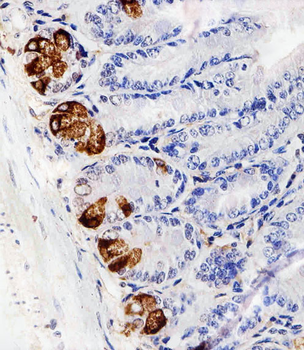
Immunohistochemical analysis of paraffin-embedded H. small intestine section using LYZ Antibody (C-term). Diluted at 1: 100 dilution. A peroxidase-conjugated goat anti-rabbit IgG at 1:400 dilution was used as the secondary antibody, followed by DAB staining.
Western blot analysis of lysates from HepG2, THP-1, WiDr cell line and human spleen tissue lysate (from left to right), using LYZ Antibody (C-term). Diluted at 1:1000 at each lane. A goat anti-rabbit IgG H&L (HRP) at 1:10000 dilution was used as the secondary antibody.
All lanes: Anti-LYZ Antibody (C-term) at 1:2000 dilution. Lane 1: THP-1 whole cell lysate. Lane 2: HepG2 whole cell lysate. Lane 3: Human spleen lysate. Lysates/proteins at 20 µg per lane. Secondary Goat Anti-Rabbit IgG, (H+L), Peroxidase conjugated at 1/10000 dilution. Predicted band size: 15 kDa. Blocking/Dilution buffer: 5% NFDM/TBST.
All lanes: Anti-LYZ Antibody (C-term) at 1:2000 dilution. Lane 1: WiDr whole cell lysate. Lane 2: THP-1 whole cell lysate. Lane 3: HepG2 whole cell lysate. Lane 4: Human spleen lysate. Lysates/proteins at 20 µg per lane. Secondary Goat Anti-Rabbit IgG, (H+L), Peroxidase conjugated at 1/10000 dilution. Predicted band size: 17 kDa. Blocking/Dilution buffer: 5% NFDM/TBST.
Staining LYZ in human tonsil tissue sections by Immunohistochemistry (IHC-P - paraformaldehyde-fixed, paraffin-embedded sections). Tissue was fixed with formaldehyde and blocked with 3% BSA for 0.5 hour at room temperature, antigen retrieval was by heat mediation with a citrate buffer (pH6). Samples were incubated with primary antibody (1/25) for 1 hours at 37C. A undiluted biotinylated goat polyvalent antibody was used as the secondary antibody.
* VAT and and shipping costs not included. Errors and price changes excepted