Supplied at 0.5 mg/ml in Tris saline, 0.02% sodium azide, pH 7.3 with 0.5% bovine serum albumin. Aliquot and store at -20C. Minimize freezing and thawing.
Sequence:
SNQKTIQPPRK
Target:
CD47
Application Dilute:
ELISA: 1:128000, WB: 0.3-1 µg/ml
Application Notes:
Application Notes: This antibody is expected to recognize both reported isoforms
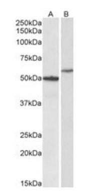
Western blot analysis of Human Hippocampus (Lane 1) and HeLa (Lane 2) lysates using CD47 antibody
Immunofluorescence analysis of paraformaldehyde fixed A431 cells, permeabilized with 0.15% Triton. Primary incubation 1 hr (10 µg/mL) followed by Alexa Fluor 488 secondary antibody (2 µg/mL), showing cytoplasmic and membrane staining. The nuclear stain is DAPI (blue). Negative control: Unimmunized goat IgG (10 µg/mL) followed by Alexa Fluor 488 secondary antibody (2 µg/mL).
Immunofluorescence analysis of paraformaldehyde fixed U2OS cells, permeabilized with 0.15% Triton. Primary incubation 1 hr (10 µg/mL) followed by Alexa Fluor 488 secondary antibody (2 µg/mL), showing membrane, cytoplasmic and some nuclear staining. The nuclear stain is DAPI (blue). Negative control: Unimmunized goat IgG (10 µg/mL) followed by Alexa Fluor 488 secondary antibody (2 µg/mL).
7 µg/mL staining of paraffin embedded Human Cortex. Heat induced antigen retrieval with citrate buffer pH 6, HRP-staining.
Negative Control showing staining of paraffin embedded Human Cortex, with no primary antibody.
Immunofluorescence analysis of paraformaldehyde fixed THP-1 cells immobilized on ShifixTM coverslip, permeabilized with 0.15% Triton. Primary incubation 1 hr (10 µg/mL) followed by Alexa Fluor 488 secondary antibody (2 µg/mL), showing membrane and cytoplasmic staining. The nuclear stain is DAPI (blue). Negative control: Unimmunized goat IgG (10 µg/mL) followed by Alexa Fluor 488 secondary antibody (2 µg/mL).
* VAT and and shipping costs not included. Errors and price changes excepted