WB (1:1000), IHC (1:1000), ICC/IF (1:200), optimal dilutions for assays should be determined by the user.
Application Notes:
Application Notes: 1 µg/ml of SMC-118 was sufficient for detection of HSF1 in 20 µg of heat shocked HeLa cell lysate by ECL immunoblot analysis using Goat anti-rat IgG: HRP as the secondary antibody
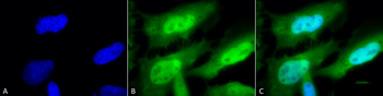
Immunocytochemistry/Immunofluorescence analysis using Rat Anti-HSF1 Monoclonal Antibody, Clone 10H8. Tissue: Heat Shocked cervical cancer cells (HeLa). Species: Human. Fixation: 2% Formaldehyde for 20 min at RT. Primary Antibody: Rat Anti-HSF1 Monoclonal Antibody at 1:100 for 12 hours at 4C. Secondary Antibody: FITC Goat Anti-Rat (green) at 1:200 for 2 hours at RT. Counterstain: DAPI (blue) nuclear stain at 1:40000 for 2 hours at RT. Localization: Diffuse nuclear and cytoplasmic staining. Magnification: 100x. (A) DAPI (blue) nuclear stain. (B) Anti-HSF1 Antibody. (C) Composite. Heat Shocked at 42C for 1h.
Immunohistochemistry analysis using Rat Anti-HSF1 Monoclonal Antibody, Clone 10H8. Tissue: Breast carcinoma. Species: Human. Fixation: 10% Formalin Solution for 20 hours at RT. Primary Antibody: Rat Anti-HSF1 Monoclonal Antibody at 1:1000 for 40 min. Secondary Antibody: Dako labeled Polymer HRP Anti-rat IgG, DAB Chromogen (brown) (Dako Envision+ System) for 30 min at RT. Counterstain: Mayers Hematoxylin (purple/blue) nuclear stain for 1 minute at RT. Localization: Nuclear. Magnification: 100X.
Immunocytochemistry/Immunofluorescence analysis using Rat Anti-HSF1 Monoclonal Antibody, Clone 10H8. Tissue: Heat Shocked mitotic HeLa cells. Species: Human. Primary Antibody: Rat Anti-HSF1 Monoclonal Antibody at 1:1000. HSF1 stained green.
Western Blot analysis of Human Breast adenocarcinoma cell line (MCF7) showing detection of ~65 kDa HSF1 protein using Rat Anti-HSF1 Monoclonal Antibody, Clone 10H8. Lane 1: MW ladder. Lane 2: HSF1 null lysate prepared from mouse embryonic fibroblasts. Lane 3: MCF7 lysate (5 µg). Lane 4: MCF7 lysate (10 µg). Lane 5: MCF7 lysate (20 µg). Block: 1.5% BSA for 30 minutes at RT. Primary Antibody: Rat Anti-HSF1 Monoclonal Antibody at 1:1000 for 2 hours at RT. Secondary Antibody: Goat Anti-Rat IgG: HRP for 1 hour at RT. Predicted/Observed Size: ~65 kDa.
Immunocytochemistry/Immunofluorescence analysis using Rat Anti-HSF1 Monoclonal Antibody, Clone 10H8. Tissue: Heat Shocked cervical cancer cells (HeLa). Species: Human. Fixation: 2% Formaldehyde for 20 min at RT. Primary Antibody: Rat Anti-HSF1 Monoclonal Antibody at 1:100 for 12 hours at 4C. Secondary Antibody: APC Goat Anti-Rat (red) at 1:200 for 2 hours at RT. Counterstain: DAPI (blue) nuclear stain at 1:40000 for 2 hours at RT. Localization: Diffuse nuclear and cytoplasmic staining. Magnification: 20x. (A) DAPI (blue) nuclear stain. (B) Anti-HSF1 Antibody. (C) Composite. Heat Shocked at 42C for 1h.
Immunohistochemistry analysis using Rat Anti-HSF1 Monoclonal Antibody, Clone 10H8. Tissue: Lung. Species: Mouse. Fixation: 10% Formalin Solution for 20 hours at RT. Primary Antibody: Rat Anti-HSF1 Monoclonal Antibody at 1:1000 for 40 min. Secondary Antibody: Dako labeled Polymer HRP Anti-rat IgG, DAB Chromogen (brown) (Dako Envision+ System) for 30 min at RT. Counterstain: Mayers Hematoxylin (purple/blue) nuclear stain for 1 minute at RT. Localization: Nuclear. Magnification: 100X. (A) HSF Wildtype. (B) HSF null.
* VAT and and shipping costs not included. Errors and price changes excepted