WB (1:1000), IHC (1:200), ICC/IF (1:200), optimal dilutions for assays should be determined by the user.
Application Notes:
Application Notes: 1 µg/ml of SMC-106 was sufficient for detection of HSP70 and HSC70 in 20 µg of heat shocked HeLa cell lysate by colorimetric immunoblot analysis using Goat anti-mouse IgG:HRP as the secondary antibody
Immunohistochemistry analysis using Mouse Anti-Hsp70 Monoclonal Antibody, Clone BB70. Tissue: inflamed colon. Species: Mouse. Fixation: Formalin. Primary Antibody: Mouse Anti-Hsp70 Monoclonal Antibody at 1:10000 for 12 hours at 4C. Secondary Antibody: Biotin Goat Anti-Mouse at 1:2000 for 1 hour at RT. Counterstain: Mayer Hematoxylin (purple/blue) nuclear stain at 200 µl for 2 minutes at RT. Localization: Inflammatory cells. Magnification: 40x. Inflammatory cells. HSP70/HSC70 stained brown.
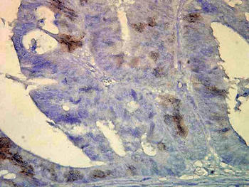
Immunohistochemistry analysis using Mouse Anti-Hsp70 Monoclonal Antibody, Clone BB70. Tissue: colon carcinoma. Species: Human. Fixation: Formalin. Primary Antibody: Mouse Anti-Hsp70 Monoclonal Antibody at 1:10000 for 12 hours at 4C. Secondary Antibody: Biotin Goat Anti-Mouse at 1:2000 for 1 hour at RT. Counterstain: Mayer Hematoxylin (purple/blue) nuclear stain at 200 µl for 2 minutes at RT. Localization: Inflammatory cells. Magnification: 40x. HSP70/HSC70 cells stained brown.
Western Blot analysis of Bovine MDBK cell lysates showing detection of Hsp70 protein using Mouse Anti-Hsp70 Monoclonal Antibody, Clone BB70. Primary Antibody: Mouse Anti-Hsp70 Monoclonal Antibody at 1:1000.
Western Blot analysis of Human Cervical cancer cell line (HeLa) lysate showing detection of Hsp70 protein using Mouse Anti-Hsp70 Monoclonal Antibody, Clone BB70. Primary Antibody: Mouse Anti-Hsp70 Monoclonal Antibody at 1:1000. Secondary Antibody: HRP Goat Anti-Rat.
Immunohistochemistry analysis using Mouse Anti-Hsp70 Monoclonal Antibody, Clone BB70. Tissue: hepatocytes. Species: Rat. Fixation: Paraffin Embedded. Primary Antibody: Mouse Anti-Hsp70 Monoclonal Antibody at 1:200. Liver sections were paraffin embedded. First pictures in series show two hours after exposure to stress, the second shows the control.
Immunocytochemistry/Immunofluorescence analysis using Mouse Anti-Hsp70 Monoclonal Antibody, Clone BB70. Tissue: hepatocyte nuclei. Species: Rat. Primary Antibody: Mouse Anti-Hsp70 Monoclonal Antibody at 1:200. Liver sections were paraffin embedded. First pictures in series show two hours after exposure to stress, the second shows the control.
* VAT and and shipping costs not included. Errors and price changes excepted