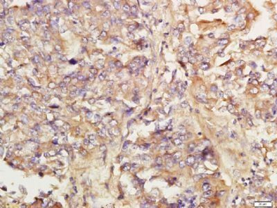
Immunohistochemical staining of human lung carcinoma tissue using KIAA0701 antibody.
Immunohistochemical staining of mouse embryo tissue using KIAA0701 antibody.
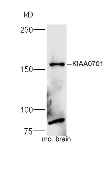
Protein: brain (mouse) lysate at 40 ug, Primary: rabbit Anti-KIAA0701 (orb184706) at 1:300, Secondary: HRP conjugated Goat-Anti-rabbit IgG (orb572747) at 1:5000, Predicted band size: 161 kD, Observed band size: 161kD.
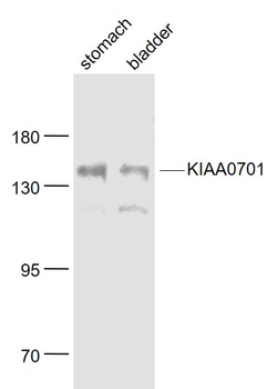
Sample: Stomach (Mouse) Lysate at 40 ug, Bladder (Mouse) Lysate at 40 ug, Primary: Anti-KIAA0701 (orb184706) at 1/1000 dilution, Secondary: IRDye800CW Goat Anti-Rabbit IgG at 1/20000 dilution, Predicted band size: 161 kD, Observed band size: 161 kD.
Tissue/Cell: human lung carcinoma, 4% Paraformaldehyde-fixed and paraffin-embedded, Antigen retrieval: citrate buffer (0.01M, pH 6.0), Boiling bathing for 15 min, Block endogenous peroxidase by 3% Hydrogen peroxide for 30 min, Blocking buffer (normal goat serum) at 37C for 20 min, Incubation: Anti-KIAA0701 Polyclonal Antibody, Unconjugated (orb184706) 1:200, overnight at 4C, followed by conjugation to the secondary antibody and DAB staining.
* VAT and and shipping costs not included. Errors and price changes excepted