A synthetic peptide corresponding to a sequence at the C-terminus of human Bag1, different from the related mouse sequence by two amino acids, and from the related rat sequence by three amino acids.
Conjugation:
Unconjugated
Alternative Names:
BAG family molecular chaperone regulator 1, BAG-1, Bcl-2-associated athanogene 1, BAG1, HAP
Bag1 Rabbit Polyclonal Antibody
Clonality:
Polyclonal
Concentration:
Adding 0.2 ml of distilled water will yield a concentration of 500 µg/ml.
Each vial contains 4mg Trehalose, 0.9mg NaCl and 0.2mg Na2HPO4.
Form:
Lyophilized
Target:
BAG family molecular chaperone regulator 1
Application Dilute:
Western blot, 0.1-0.5µg/ml, Human Immunohistochemistry (Paraffin-embedded Section), 2-5µg/ml, Human, Mouse, Rat
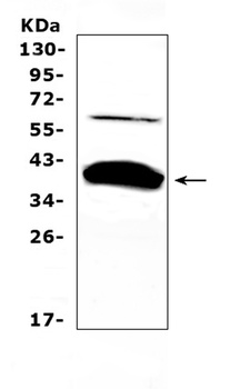
WB analysis of Bag1 using anti-Bag1 antibody.Lane 1:rat kidney tissue.
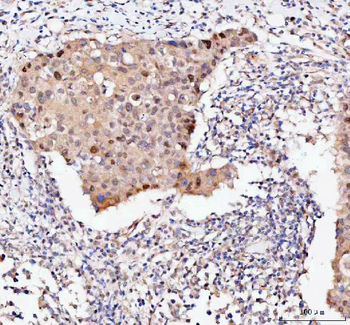
IHC analysis of Bag1 using anti-Bag1 antibody. Bag1 was detected in a paraffin-embedded section of human breast cancer tissue. Heat mediated antigen retrieval was performed in EDTA buffer (pH8.0, epitope retrieval solution). The tissue section was blocked with 10% goat serum. The tissue section was then incubated with 2 µg/ml rabbit anti-Bag1 Antibody overnight at 4C. Peroxidase Conjugated Goat Anti-rabbit IgG was used as secondary antibody and incubated for 30 minutes at 37C. The tissue section was developed using HRP Conjugated Rabbit IgG Super Vision Assay Kit with DAB as the chromogen.
IHC analysis of Bag1 using anti-Bag1 antibody. Bag1 was detected in a paraffin-embedded section of human colorectal adenocarcinoma tissue. Heat mediated antigen retrieval was performed in EDTA buffer (pH8.0, epitope retrieval solution). The tissue section was blocked with 10% goat serum. The tissue section was then incubated with 2 µg/ml rabbit anti-Bag1 Antibody overnight at 4C. Peroxidase Conjugated Goat Anti-rabbit IgG was used as secondary antibody and incubated for 30 minutes at 37C. The tissue section was developed using HRP Conjugated Rabbit IgG Super Vision Assay Kit with DAB as the chromogen.
IHC analysis of Bag1 using anti-Bag1 antibody. Bag1 was detected in a paraffin-embedded section of human tonsil tissue. Heat mediated antigen retrieval was performed in EDTA buffer (pH8.0, epitope retrieval solution). The tissue section was blocked with 10% goat serum. The tissue section was then incubated with 2 µg/ml rabbit anti-Bag1 Antibody overnight at 4C. Peroxidase Conjugated Goat Anti-rabbit IgG was used as secondary antibody and incubated for 30 minutes at 37C. The tissue section was developed using HRP Conjugated Rabbit IgG Super Vision Assay Kit with DAB as the chromogen.
IHC analysis of Bag1 using anti-Bag1 antibody. Bag1 was detected in a paraffin-embedded section of mouse colon tissue. Heat mediated antigen retrieval was performed in EDTA buffer (pH8.0, epitope retrieval solution). The tissue section was blocked with 10% goat serum. The tissue section was then incubated with 2 µg/ml rabbit anti-Bag1 Antibody overnight at 4C. Peroxidase Conjugated Goat Anti-rabbit IgG was used as secondary antibody and incubated for 30 minutes at 37C. The tissue section was developed using HRP Conjugated Rabbit IgG Super Vision Assay Kit with DAB as the chromogen.
IHC analysis of Bag1 using anti-Bag1 antibody. Bag1 was detected in a paraffin-embedded section of rat colon tissue. Heat mediated antigen retrieval was performed in EDTA buffer (pH8.0, epitope retrieval solution). The tissue section was blocked with 10% goat serum. The tissue section was then incubated with 2 µg/ml rabbit anti-Bag1 Antibody overnight at 4C. Peroxidase Conjugated Goat Anti-rabbit IgG was used as secondary antibody and incubated for 30 minutes at 37C. The tissue section was developed using HRP Conjugated Rabbit IgG Super Vision Assay Kit with DAB as the chromogen.
Western blot analysis of Bag1 using anti-Bag1 antibody. Electrophoresis was performed on a 5-20% SDS-PAGE gel at 70V (Stacking gel) / 90V (Resolving gel) for 2-3 hours. The sample well of each lane was loaded with 30 ug of sample under reducing conditions. Lane 1: human Hela whole cell lysates, Lane 2: human Jurkat whole cell lysates, Lane 3: human MCF-7 whole cell lysates, Lane 4: human 293T whole cell lysates. After electrophoresis, proteins were transferred to a nitrocellulose membrane at 150 mA for 50-90 minutes. Blocked the membrane with 5% non-fat milk/TBS for 1.5 hour at RT. The membrane was incubated with rabbit anti-Bag1 antigen affinity purified polyclonal antibody at 0.5 µg/mL overnight at 4C, then washed with TBS-0.1% Tween 3 times with 5 minutes each and probed with a goat anti-rabbit IgG-HRP secondary antibody at a dilution of 1:5000 for 1.5 hour at RT. The signal is developed using an Enhanced Chemiluminescent detection (ECL) kit with Tanon 5200 system. A specific band was detected for
* VAT and and shipping costs not included. Errors and price changes excepted