Purified polyclonal antibody supplied in PBS with 0.09% (W/V) sodium azide. This antibody is purified through a protein A column, followed by peptide affinity purification.
Application Dilute:
IF - 1:25, IHC-P - 1:100-500, WB - 1:1000
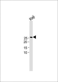
All lanes: Anti-AK4 Antibody (Center) at 1:1000 dilution + Raji whole cell lysate. Lysates/proteins at 20 µg per lane. Secondary Goat Anti-Rabbit IgG, (H+L), Peroxidase conjugated at 1/15000 dilution.Observed band size: 26kDa. Blocking/Dilution buffer: 5% NFDM/TBST.
Immunohistochemical analysis of paraffin-embedded H.heart section using AK4 Antibody (Center). Diluted at 1:25 dilution. A peroxidase-conjugated goat anti-rabbit IgG at 1:400 dilution was used as the secondary antibody, followed by DAB staining.
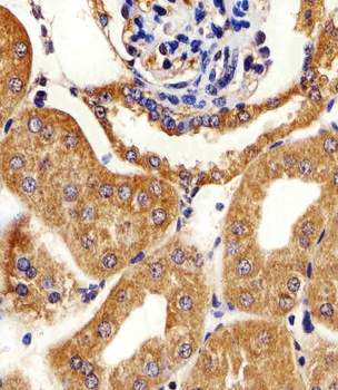
Immunohistochemical analysis of paraffin-embedded H.kidney section using AK4 Antibody (Center). Diluted at 1:25 dilution. A peroxidase-conjugated goat anti-rabbit IgG at 1:400 dilution was used as the secondary antibody, followed by DAB staining.
Immunohistochemical analysis of paraffin-embedded M.kidney section using AK4 Antibody (Center). Diluted at 1:25 dilution. A peroxidase-conjugated goat anti-rabbit IgG at 1:400 dilution was used as the secondary antibody, followed by DAB staining.
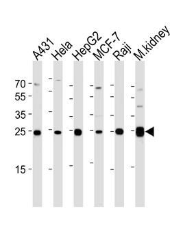
Western blot analysis of lysates from A431, Hela, HepG2, MCF-7, Raji cell line and mouse kidney tissue lysate (from left to right), using AK4 Antibody (Center). Diluted at 1:1000 at each lane. A goat anti-rabbit IgG H&L (HRP) at 1:5000 dilution was used as the secondary antibody. Lysates at 35 ug per lane.
Fluorescent image of HepG2 cells stained with AK4 Antibody (Center). Diluted at 1:25 dilution. An Alexa Fluor 488-conjugated goat anti-rabbit lgG at 1:400 dilution was used as the secondary antibody (green). DAPI was used to stain the cell nuclear (blue). Cytoplasmic actin was counterstained with Alexa Fluor 555 conjugated with Phalloidin (red).
* VAT and and shipping costs not included. Errors and price changes excepted