Purified polyclonal antibody supplied in PBS with 0.09% (W/V) sodium azide. This antibody is purified through a protein A column, followed by peptide affinity purification.
Application Dilute:
IHC-P - 1:100-500, WB - 1:1000, IF - 1:10-50, FC - 1:10-50
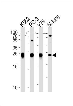
GSTP1 Antibody (Center) western blot analysis in K562, PC-3, Y79 cell line and mouse lung tissue lysates (35 ug/lane).This demonstrates the GSTP1 antibody detected the GSTP1 protein (arrow).
GSTP1 Antibody (Center) flow cytometric analysis of Ramos cells (right histogram) compared to a negative control cell (left histogram). FITC-conjugated goat-anti-rabbit secondary antibodies were used for the analysis.
Immunohistochemical analysis of paraffin-embedded Human tonsil section using Pink1. Diluted at 1:2000 dilution. A undiluted biotinylated goat polyvalent antibody was used as the secondary, followed by DAB staining.
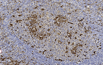
GSTP1 Antibody (Center) IHC analysis in formalin fixed and paraffin embedded human colon carcinoma followed by peroxidase conjugation of the secondary antibody and DAB staining. This data demonstrates the use of the GSTP1 Antibody (Center) for immunohistochemistry. Clinical relevance has not been evaluated.
Fluorescent confocal image of A549 cell stained with GSTP1 Antibody (Center).A549 cells were fixed with 4% PFA (20 min), permeabilized with Triton X-100 (0.1%, 10 min), then incubated with GSTP1 primary antibody (1: 25, 1 h at 37C). For secondary antibody, Alexa Fluor 488 conjugated donkey anti-rabbit antibody (green) was used (1:400, 50 min at 37C).Cytoplasmic actin was counterstained with Alexa Fluor 555 (red) conjugated Phalloidin (7units/ml, 1 h at 37C). Nuclei were counterstained with DAPI (blue) (10 µg/ml, 10 min). GSTP1 immunoreactivity is localized to Cytoplasm significantly.
Fluorescent confocal image of A549 cell stained with GSTP1 Antibody (Center).A549 cells were fixed with 4% PFA (20 min), permeabilized with Triton X-100 (0.1%, 10 min), then incubated with GSTP1 primary antibody (1: 25, 1 h at 37C). For secondary antibody, Alexa Fluor 488 conjugated donkey anti-rabbit antibody (green) was used (1:400, 50 min at 37C).Cytoplasmic actin was counterstained with Alexa Fluor 555 (red) conjugated Phalloidin (7units/ml, 1 h at 37C). Nuclei were counterstained with DAPI (blue) (10 µg/ml, 10 min). GSTP1 immunoreactivity is localized to Cytoplasm and Mitochondria significantly.
* VAT and and shipping costs not included. Errors and price changes excepted