Purified polyclonal antibody supplied in PBS with 0.09% (W/V) sodium azide. This antibody is purified through a protein A column, followed by peptide affinity purification.
Application Dilute:
WB - 1:1000
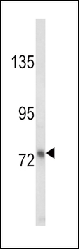
Western blot analysis of LTF Antibody in MDA-MB231 cell line lysates (35 ug/lane). LTF (arrow) was detected using the purified Pab.
Western blot analysis of LTF Antibody in mouse spleen tissue lysates (35 ug/lane). LTF (arrow) was detected using the purified Pab.
LTF Antibody flow cytometric analysis of MDA-MB231 cells (right histogram) compared to a negative control cell (left histogram). FITC-conjugated goat-anti-rabbit secondary antibodies were used for the analysis.
All lanes: Anti-LTF Antibody at 1:1000-2000 dilution. Lane 1: Human plasma tissue lysate. Lane 2: Hela whole cell lysate. Lane 3: Human uterus tissue lysate. Lysates/proteins at 20 µg per lane. Secondary Goat Anti-Rabbit IgG, (H+L), Peroxidase conjugated at 1/10000 dilution. Predicted band size: 78 kDa. Blocking/Dilution buffer: 5% NFDM/TBST.
All lanes: Anti-LTF Antibody at 1:1000 dilution. Lane 1: Human plasma tissue lysate. Lane 2: Human fetal kidney tissue lysate. Lane 3: Human uterus tissue lysate. Lane 4: Human skeletal muscle tissue lysate. Lysates/proteins at 20 µg per lane. Secondary Goat Anti-Rabbit IgG, (H+L), Peroxidase conjugated at 1/10000 dilution. Predicted band size: 78 kDa. Blocking/Dilution buffer: 5% NFDM/TBST.
All lanes: Anti-LTF Antibody at 1:4000 dilution. Lane 1: A549 whole cell lysate. Lane 2: Human plasma tissue lysate. Lane 3: Human fetal kidney tissue lysate. Lane 4: Human breast tissue lysate. Lane 5: Human uterus tissue lysate. Lysates/proteins at 20 µg per lane. Secondary Goat Anti-Rabbit IgG, (H+L), Peroxidase conjugated at 1/10000 dilution. Predicted band size: 78 kDa. Blocking/Dilution buffer: 5% NFDM/TBST.
* VAT and and shipping costs not included. Errors and price changes excepted