Purified polyclonal antibody supplied in PBS with 0.09% (W/V) sodium azide. This antibody is purified through a protein A column, followed by peptide affinity purification.
Application Dilute:
WB - 1:1000, IHC-P - 1:100-500, FC - 1:10-50
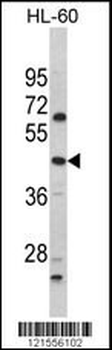
Western blot analysis of PTAR1 Antibody (Center) in HL-60 cell line lysates (35 ug/lane). PTAR1 (arrow) was detected using the purified Pab.
Western blot analysis of PTAR1 Antibody (Center) in Jurkat cell line lysates (35 ug/lane). PTAR1 (arrow) was detected using the purified Pab.
Western blot analysis of PTAR1 Antibody (Center) in MDA-MB231 cell line lysates (35 ug/lane). PTAR1 (arrow) was detected using the purified Pab.
Flow cytometric analysis of SK-Br-3 cells using PTAR1 Antibody (Center) (bottom histogram) compared to a negative control cell (top histogram). FITC-conjugated goat-anti-rabbit secondary antibodies were used for the analysis.
Anti-PTAR1 Antibody (Center) at 1:1000 dilution + CCRF-CEM whole cell lysate. Lysates/proteins at 20 µg per lane. Secondary Goat Anti-Rabbit IgG, (H+L), Peroxidase conjugated at 1/10000 dilution. Predicted band size: 46 kDa. Blocking/Dilution buffer: 5% NFDM/TBST.
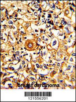
Formalin-fixed and paraffin-embedded human breast carcinoma with PTAR1 Antibody (Center), which was peroxidase-conjugated to the secondary antibody, followed by DAB staining. This data demonstrates the use of this antibody for immunohistochemistry, clinical relevance has not been evaluated.
* VAT and and shipping costs not included. Errors and price changes excepted