Purified polyclonal antibody supplied in PBS with 0.09% (W/V) sodium azide. This antibody is purified through a protein A column, followed by peptide affinity purification.
Target:
This TrkA antibody is generated from rabbits immunized with a KLH conjugated synthetic peptide between 769-796 amino acids from human TrkA.
Western blot analysis of hTrkA-pY791 in HepG2 cell line lysates (35 ug/lane). TRK (arrow) was detected using the purified Pab.
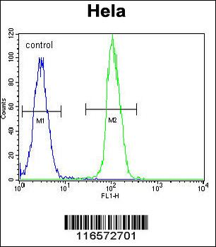
TrkA-pY791 Antibody flow cytometric analysis of Hela cells (right histogram) compared to a negative control cell (left histogram). FITC-conjugated goat-anti-rabbit secondary antibodies were used for the analysis.
Anti-TrkA (Y791) Antibody at 1:2000 dilution + mouse brain lysate. Lysates/proteins at 20 µg per lane. Secondary Goat Anti-Rabbit IgG, (H+L), Peroxidase conjugated at 1/10000 dilution. Predicted band size: 87 kDa. Blocking/Dilution buffer: 5% NFDM/TBST.
TrkA-pY791 Antibody immunohistochemistry analysis in formalin fixed and paraffin embedded human skeletal muscle followed by peroxidase conjugation of the secondary antibody and DAB staining.This data demonstrates the use of TrkA-pY791 Antibody for immunohistochemistry. Clinical relevance has not been evaluated.
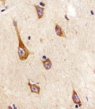
Staining TrkA in human brain tissue sections by Immunohistochemistry (IHC-P - paraformaldehyde-fixed, paraffin-embedded sections). Tissue was fixed with formaldehyde and blocked with 3% BSA for 0.5 hour at room temperature, antigen retrieval was by heat mediation with a citrate buffer (pH6). Samples were incubated with primary antibody (1/25) for 1 hours at 37C. A undiluted biotinylated goat polyvalent antibody was used as the secondary antibody.
Overlay histogram showing SH-SY5Y cells stained (green line). The cells were fixed with 2% paraformaldehyde (10 min). The cells were then icubated in 2% bovine serum albumin to block non-specific protein-protein interactions followed by the antibody (1:25 dilution) for 60 min at 37C. The secondary antibody used was Goat-Anti-Rabbit IgG, DyLight 488 Conjugated Highly Cross-Adsorbed at 1/400 dilution for 40 min at 37C. Isotype control antibody (blue line) was rabbit IgG (1 µg/1x10 6 cells) used under the same conditions. Acquisition of > 10000 events was performed.
* VAT and and shipping costs not included. Errors and price changes excepted