Purified polyclonal antibody supplied in PBS with 0.09% (W/V) sodium azide. This antibody is purified through a protein A column, followed by peptide affinity purification.
Application Dilute:
WB - 1:1000, FC - 1:10-50, IF - 1:10-50
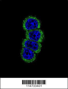
Confocal immunofluorescent analysis of ErbB2 antibody with MCF-7 cell followed by Alexa Fluor 488-conjugated goat anti-rabbit lgG (green). DAPI was used to stain the cell nuclear (blue).
Western blot analysis of HER2 (arrow) using rabbit polyclonal HER2 antibody. 293 cell lysates (2 ug/lane) either nontransfected (Lane 1) or transiently transfected with the HER2 gene (Lane 2).
Flow cytometric analysis of MCF-7 cells using HER2 Antibody (bottom histogram) compared to a negative control cell (top histogram). FITC-conjugated goat-anti-rabbit secondary antibodies were used for the analysis.
Anti-HER2 Antibody at 1:1000 dilution + SK-BR-3 whole cell lysate. Lysates/proteins at 20 µg per lane. Secondary Goat Anti-Rabbit IgG, (H+L), Peroxidase conjugated at 1/10000 dilution. Predicted band size: 138 kDa. Blocking/Dilution buffer: 5% NFDM/TBST.
All lanes: Anti-HER2/ERBB2 Antibody at 1:1000 dilution. Lane 1: SK-BR-3 whole cell lysate. Lane 2: MDA-MB-453 whole cell lysate. Lysates/proteins at 20 µg per lane. Secondary Goat Anti-Rabbit IgG, (H+L), Peroxidase conjugated at 1/10000 dilution. Predicted band size: 138 kDa. Blocking/Dilution buffer: 5% NFDM/TBST.
Immunohistochemical analysis of paraffin-embedded Human breast carcinoma tissue was performed on the Leica BOND RXm. Tissue was fixed with formaldehyde at room temperature, antigen retrieval was by heat mediation with a EDTA buffer (pH9.0). Samples were incubated with primary antibody (1:500) for 1 hours at room temperature. A undiluted biotinylated CRF Anti-Polyvalent HRP Polymer antibody was used as the secondary antibody.
* VAT and and shipping costs not included. Errors and price changes excepted