Purified polyclonal antibody supplied in PBS with 0.09% (W/V) sodium azide. This antibody is purified through a protein A column, followed by peptide affinity purification.
Application Dilute:
IHC-P - 1:100-500, WB - 1:1000
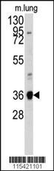
Western blot analysis of anti-ANXA2 Antibody (N-term) in mouse lung tissue lysates (35 ug/lane). ANXA2 (arrow) was detected using the purified Pab.
ANXA2 Antibody (N-term) western blot analysis in 293, MDA-MB231 cell line lysates (35 ug/lane).This demonstrates the ANXA2 antibody detected the ANXA2 protein (arrow).
Western blot analysis of ANXA2 (arrow) using rabbit polyclonal ANXA2 Antibody (N-term). 293 cell lysates (2 ug/lane) either nontransfected (Lane 1) or transiently transfected (Lane 2) with the ANXA2 gene.
Formalin-fixed and paraffin-embedded human lung carcinoma tissue reacted with ANXA2 antibody (N-term), which was peroxidase-conjugated to the secondary antibody, followed by DAB staining. This data demonstrates the use of this antibody for immunohistochemistry, clinical relevance has not been evaluated.
All lanes: Anti-ANXA2 Antibody (N-term) at 1:2000 dilution. Lane 1: Hela whole cell lysates. Lane 2: MCF-7 whole cell lysates. Lane 3: NIH/3T3 whole cell lysates. Lane 4: SH-SY5Y whole cell lysates. Lane 5: SK-BR-3 whole cell lysates. Lysates/proteins at 20 µg per lane. Secondary Goat Anti-Rabbit IgG, (H+L), Peroxidase conjugated at 1/10000 dilution. Predicted band size: 39 kDa. Blocking/Dilution buffer: 5% NFDM/TBST.
Staining ANXA2 in Human liver tissue sections by Immunohistochemistry (IHC-P - paraformaldehyde-fixed, paraffin-embedded sections). Tissue was fixed with formaldehyde and blocked with 3% BSA for 0.5 hour at room temperature, antigen retrieval was by heat mediation with a citrate buffer (pH6). Samples were incubated with primary antibody (1/25) for 1 hours at 37C. A undiluted biotinylated goat polyvalent antibody was used as the secondary antibody.
* VAT and and shipping costs not included. Errors and price changes excepted