Purified polyclonal antibody supplied in PBS with 0.09% (W/V) sodium azide. This antibody is purified through a protein A column, followed by peptide affinity purification.
Application Dilute:
IF - 1:25, FC - 1:25, WB - 1:1000
Immunohistochemical analysis of paraffin-embedded human kidney tissue was performed on the Leica BOND RXm. Samples were incubated with primary antibody (1/500) for 1 hours at room temperature. A undiluted biotinylated CRF Anti-Polyvalent HRP Polymer antibody was used as the secondary antibody.
Immunohistochemical analysis on paraffin-embedded Human liver tissue. Tissue was fixed with formaldehyde at room temperature. Heat induced epitope retrieval was performed by EDTA buffer (pH9.0). Samples were incubated with primary antibody (1:100) for 1 hour at room temperature. Undiluted CRF Anti-Polyvalent HRP Polymer antibody was used as the secondary antibody.
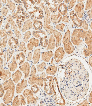
Immunohistochemical analysis on paraffin-embedded Human kidney tissue. Tissue was fixed with formaldehyde at room temperature. Heat induced epitope retrieval was performed by EDTA buffer (pH9.0). Samples were incubated with primary antibody (1:100) for 1 hour at room temperature. Undiluted CRF Anti-Polyvalent HRP Polymer antibody was used as the secondary antibody.
Immunofluorescent analysis of 4% paraformaldehyde-fixed, 0.1% Triton X-100 permeabilized U-2OS cells labeling RPS18 at 1/25 dilution, followed by Dylight 488-conjugated goat anti-Rabbit IgG secondary antibody at 1/200 dilution (green). Immunofluorescence image showing cytoplasm staining on U-2OS cell line. Cytoplasmic actin is detected with Dylight 554 Phalloidin (1186255) at 1/500 dilution (red). The nuclear counter stain is DAPI (blue).
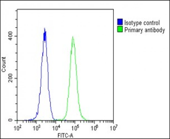
Overlay histogram showing U-2 OS cells stained (green line). The cells were fixed with 2% paraformaldehyde (10 min) and then permeabilized with 90% methanol for 10 min. The cells were then icubated in 2% bovine serum albumin to block non-specific protein-protein interactions followed by the antibody (1:25 dilution) for 60 min at 37C. The secondary antibody used was Goat-Anti-Rabbit IgG, DyLight 488 Conjugated Highly Cross-Adsorbed at 1/200 dilution for 40 min at 37C. Isotype control antibody (blue line) was rabbit IgG1 (1 µg/1x10 6 cells) used under the same conditions. Acquisition of > 10000 events was performed.
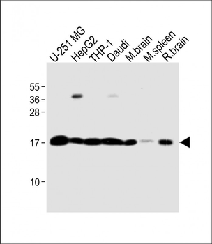
All lanes: Anti-RS18 Antibody (Center) at 1:1000 dilution. Lane 1: U-251 MG whole cell lysate. Lane 2: HepG2 whole cell lysate. Lane 3: THP-1 whole cell lysate. Lane 4: Daudi whole cell lysate. Lane 5: Mouse brain lysate. Lane 6: Mouse spleen lysate. Lane 7: Rat brain lysate. Lysates/proteins at 20 µg per lane. Secondary Goat Anti-Rabbit IgG, (H+L), Peroxidase conjugated at 1/10000 dilution. Predicted band size: 18 kDa. Blocking/Dilution buffer: 5% NFDM/TBST.
* VAT and and shipping costs not included. Errors and price changes excepted