Purified polyclonal antibody supplied in PBS with 0.09% (W/V) sodium azide. This antibody is purified through a protein A column, followed by peptide affinity purification.
Application Dilute:
FC - 1:25, IHC-P - 1:100-500, WB - 1:2000
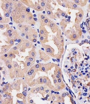
Staining Villin-1 in human kidney tissue sections by Immunohistochemistry (IHC-P - paraformaldehyde-fixed, paraffin-embedded sections). Tissue was fixed with formaldehyde and blocked with 3% BSA for 0.5 hour at room temperature, antigen retrieval was by heat mediation with a citrate buffer (pH6). Samples were incubated with primary antibody (1/25) for 1 hours at 37C. A undiluted biotinylated goat polyvalent antibody was used as the secondary antibody.
Overlay histogram showing Hela cells stained (green line). The cells were fixed with 2% paraformaldehyde (10 min) and then permeabilized with 90% methanol for 10 min. The cells were then icubated in 2% bovine serum albumin to block non-specific protein-protein interactions followed by the antibody (1:25 dilution) for 60 min at 37C. The secondary antibody used was Goat-Anti-Rabbit IgG, DyLight 488 Conjugated Highly Cross-Adsorbed at 1/200 dilution for 40 min at 37C. Isotype control antibody (blue line) was rabbit IgG1 (1 µg/1x10 6 cells) used under the same conditions. Acquisition of > 10000 events was performed.
All lanes: Anti-Villin-1 Antibody (N-term) at 1:1000 dilution. Lane 1: Caco2 whole cell lysate. Lane 2: HT-29 whole cell lysate. Lysates/proteins at 20 µg per lane. Secondary Goat Anti-Rabbit IgG, (H+L), Peroxidase conjugated at 1/10000 dilution. Predicted band size: 93 kDa. Blocking/Dilution buffer: 5% NFDM/TBST.
All lanes: Anti-Villin-1 Antibody (N-term) at 1: 8000 dilution. Lane 1: HT-29 whole cell lysate. Lane 2: SW480 whole cell lysate. Lane 3: Mouse colon lysate. Lysates/proteins at 20 µg per lane. Secondary Goat Anti-Rabbit IgG, (H+L), Peroxidase conjugated at 1/10000 dilution. Predicted band size: 93 kDa. Blocking/Dilution buffer: 5% NFDM/TBST.
All lanes: Anti-Villin-1 Antibody (N-term) at 1:2000 dilution. Lane 1: HepG2 whole cell lysate. Lane 2: HT-29 whole cell lysate. Lane 3: Mouse kidney lysate. Lysates/proteins at 20 µg per lane. Secondary Goat Anti-Rabbit IgG, (H+L), Peroxidase conjugated at 1/10000 dilution. Predicted band size: 93 kDa. Blocking/Dilution buffer: 5% NFDM/TBST.
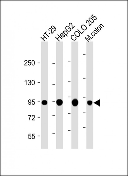
All lanes: Anti-Villin-1 Antibody (N-term) at 1:2000 dilution. Lane 1: HT-29 whole cell lysate. Lane 2: HepG2 whole cell lysate. Lane 3: COLO 205 whole cell lysate. Lane 4: Mouse colon lysate. Lysates/proteins at 20 µg per lane. Secondary Goat Anti-Rabbit IgG, (H+L), Peroxidase conjugated at 1/10000 dilution. Predicted band size: 93 kDa. Blocking/Dilution buffer: 5% NFDM/TBST.
* VAT and and shipping costs not included. Errors and price changes excepted