Purified polyclonal antibody supplied in PBS with 0.05% (V/V) Proclin 300. This antibody is prepared by Saturated Ammonium Sulfate (SAS) precipitation followed by dialysis against PBS.
Application Dilute:
WB - 1:2000, IF - 1:25, IHC-P - 1:100-500
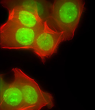
Immunofluorescent analysis of 4% paraformaldehyde-fixed, 0.1% Triton X-100 permeabilized MCF-7 (human breast cancer cell line) cells labeling CUX1 at 1/25 dilution, followed by Dylight 488-conjugated goat anti-rabbit IgG secondary antibody at 1/200 dilution (green). Immunofluorescence image showing nucleus and weak cytoplasm staining on MCF-7 cell line. Cytoplasmic actin is detected with Dylight 554 Phalloidin at 1/100 dilution (red).
Staining CUX1 in human kidney tissue sections by Immunohistochemistry (IHC-P - paraformaldehyde-fixed, paraffin-embedded sections). Tissue was fixed with formaldehyde and blocked with 3% BSA for 0.5 hour at room temperature, antigen retrieval was by heat mediation with a citrate buffer (pH6). Samples were incubated with primary antibody (1/25) for 1 hours at 37C. A undiluted biotinylated goat polyvalent antibody was used as the secondary antibody.
Staining CUX1 in human lymph node tissue sections by Immunohistochemistry (IHC-P - paraformaldehyde-fixed, paraffin-embedded sections). Tissue was fixed with formaldehyde and blocked with 3% BSA for 0.5 hour at room temperature, antigen retrieval was by heat mediation with a citrate buffer (pH6). Samples were incubated with primary antibody (1/25) for 1 hours at 37C. A undiluted biotinylated goat polyvalent antibody was used as the secondary antibody.
All lanes: Anti-CUX1 Antibody (C-term) at 1:2000 dilution. Lane 1: 293 whole cell lysate. Lane 2: K562 whole cell lysate. Lysates/proteins at 20 µg per lane. Secondary Goat Anti-Rabbit IgG, (H+L), Peroxidase conjugated at 1/10000 dilution. Predicted band size: 164 kDa. Blocking/Dilution buffer: 5% NFDM/TBST.
All lanes: Anti-CUX1 Antibody (C-term) at 1:2000 dilution. Lane 1: MCF-7 whole cell lysate. Lane 2: MOLT-4 whole cell lysate. Lane 3: SH-SY5Y whole cell lysate. Lysates/proteins at 20 µg per lane. Secondary Goat Anti-Rabbit IgG, (H+L), Peroxidase conjugated at 1/10000 dilution. Predicted band size: 164 kDa. Blocking/Dilution buffer: 5% NFDM/TBST.
All lanes: Anti-CUX1 Antibody (C-term) at 1:1000 dilution. Lane 1: SH-SY5Y whole cell lysate. Lane 2: SK-BR-3 whole cell lysate. Lane 3: NIH/3T3 whole cell lysate. Lysates/proteins at 20 µg per lane. Secondary: Goat Anti-Rabbit IgG, (H+L), Peroxidase conjugated at 1/15000 dilution. Observed band size: 200 KDa. Blocking/Dilution buffer: 5% NFDM/TBST.
* VAT and and shipping costs not included. Errors and price changes excepted