Purified polyclonal antibody supplied in PBS with 0.09% (W/V) sodium azide. This antibody is prepared by Saturated Ammonium Sulfate (SAS) precipitation followed by dialysis against PBS.
Application Dilute:
WB - 1:1000, IF - 1:10-50, IHC-P - 1:100-500, FC - 1:10-50
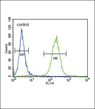
ATP1A2 Antibody (Center) flow cytometric analysis of MCF-7 cells (right histogram) compared to a negative control cell (left histogram). FITC-conjugated goat-anti-rabbit secondary antibodies were used for the analysis.
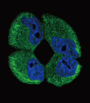
Confocal immunofluorescent analysis of ATP1A2 Antibody (Center) with MCF-7 cell followed by Alexa Fluor488-conjugated goat anti-rabbit lgG (green). DAPI was used to stain the cell nuclear (blue).
ATP1A2 Antibody (Center) immunohistochemistry analysis in formalin fixed and paraffin embedded human brain tissue followed by peroxidase conjugation of the secondary antibody and DAB staining. This data demonstrates the use of the ATP1A2 Antibody (Center) for immunohistochemistry. Clinical relevance has not been evaluated.
ATP1A2 Antibody (Center) western blot analysis in MCF-7 cell line lysates (15 ug/lane). This demonstrates the ATP1A2 antibody detected ATP1A2 protein (arrow).
ATP1A2 Antibody (Center) western blot analysis in mouse heart tissue lysates (15 ug/lane). This demonstrates the ATP1A2 antibody detected ATP1A2 protein (arrow).
Anti-ATP1A2 Antibody (Center) at 1:2000 dilution + human skeletal muscle lysate. Lysates/proteins at 20 µg per lane. Secondary Goat Anti-Rabbit IgG, (H+L), Peroxidase conjugated at 1/10000 dilution. Predicted band size: 112 kDa. Blocking/Dilution buffer: 5% NFDM/TBST.
* VAT and and shipping costs not included. Errors and price changes excepted