Purified GST-p27 fusion protein of human origin was used as the immunogen for this p27Kip1 antibody.
Conjugation:
Unconjugated
This antibody recognizes a 27kDa protein, identified as the p27Kip1, a cell cycle regulatory mitotic inhibitor. The p27Kip1 antibody is highly specific and shows no cross-reaction with other related mitotic inhibitors. p27Kip1 functions as a negative regulator of G1 progression and has been proposed to function as a possible mediator of TGF beta induced G1 arrest. It is a candidate tumor suppressor gene. This mAb is excellent for staining of formalin-fixed tissues.
Clonality:
Monoclonal
Clone Designation:
[SX53G8]
Buffer:
0.2 mg/ml in 1X PBS with 0.1 mg/ml rAlbumin and 0.05% sodium azide
Application Dilute:
Flow cytometry: 0.5-1ug/million cells,Immunofluorescence: 0.5-1ug/ml,Immunohistochemistry (FFPE): 0.25-0.5ug/ml for 30 min at RT
Application Notes:
Application Notes: The concentration stated for each application is a general starting point. Variations in protocols, secondaries and substrates may require the p27Kip1 antibody to be titered up or down for optimal performance.1. Staining of formalin-fixed tissues requires boiling tissue sections in 10mM citrate buffer, pH 6.0, for 10-20 min followed by cooling at RT for 20 minutes.2. The prediluted format is supplied in a dropper bottle and is optimized for use in IHC. After epitope retrieval step (if required), drip mAb solution onto the tissue section and incubate at RT for 30 min
Immunofluorescent staining of PFA-fixed human MCF7 cells with p27Kip1 antibody (clone SX53G8, green) and Phalloidin (red).
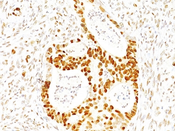
IHC staining of FFPE human prostate carcinoma tissue with p287Kip1 antibody (clone SX53G8). HIER: boil tissue sections in pH8 EDTA for 20 min and allow to cool before testing.
IHC staining of FFPE human colon carcinoma tissue with p287Kip1 antibody (clone SX53G8). HIER: boil tissue sections in pH8 EDTA for 20 min and allow to cool before testing.
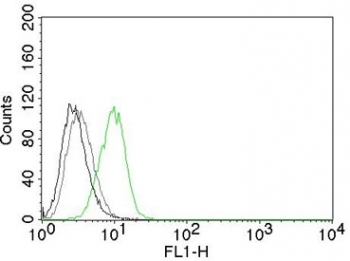
Intracellular FACS testing of human HeLa cells with Alexa Fluor 488 conjugated p27Kip1 antibody (clone SX53G8, green) and isotype control (gray).
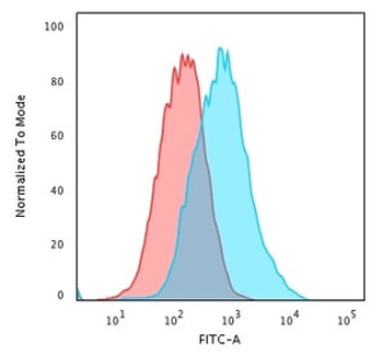
Flow cytometry staining of PFA-fixed human MCF7 cells with p27Kip1 antibody, Red = isotype control, Blue = p27Kip1 antibody.
SDS-PAGE analysis of purified, BSA-free p27Kip1 antibody (clone SX53G8) as confirmation of integrity and purity.
* VAT and and shipping costs not included. Errors and price changes excepted