The extract of pigmented melanoma metastases from lymph nodes was used as the immunogen for this gp100 antibody.
Conjugation:
Unconjugated
Melanocytes produce organelles called melanosomes which produce melanin, the pigment that gives color to skin, hair, eyes, scales and feathers. gp100 was identified in an attempt to clone the gene Tyrosinase, an enzyme required for melanin synthesis. Further testing determined that gp100 is a melanoma-specific protein and is responsible for melanosome maturation, facilitating the transition from amorphous rounded vesicles to fibrillary ellipsoid organelles.Metastatic amelanotic melanoma can often be confused with a variety of poorly differentiated carcinomas, large cell lymphomas, and sarcomas using H & E stains alone. It is also difficult to differentiate melanoma from spindle cell carcinomas and various types of mesenchymal neoplasms. Clone HMB45 gp100 antibody stains fetal and neonatal melanocytes, junctional and blue nevus cells, and malignant melanoma. It also stains angiomyolipomas, tumors most commonly associated with the kidney. Intradermal nevi, normal adult melanocytes, and non-melanocytic cells are negative. This gp100 antibody does not stain tumor cells of epithelial, lymphoid, glial, or mesenchymal origin.The gp100 molecule is a 100kDa glycosylated protein that is cleaved into a small (26kDa) carboxy-terminal fragment and a larger amino- terminal section (60-64 kDa), which is subsequently cleaved to generate 26kDa and 34-38kDa fragments.
Clonality:
Monoclonal
Clone Designation:
[HMB45]
Buffer:
0.2 mg/ml in 1X PBS with 0.1 mg/ml rAlbumin and 0.05% sodium azide
Application Dilute:
Western blot: 1-2ug/ml,Flow cytometry: 0.5-1ug/10 6 cells,Immunofluorescence: 0.5-1ug/ml,Immunohistochemistry (FFPE): 0.5-1ug/ml for 30 min at RT (1)
Application Notes:
Application Notes: The concentration stated for each application is a general starting point. Variations in protocols, secondaries and substrates may require the gp100 antibody to be titered up or down for optimal performance.1. The prediluted format is supplied in a dropper bottle and is optimized for use in IHC. After epitope retrieval step (if required), drip mAb solution onto the tissue section and incubate at RT for 30 min
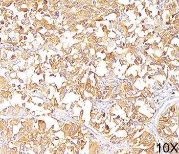
IHC staining of human melanoma (10X) with gp100 antibody (clone HMB45). HIER: boil tissue sections in pH9 10mM Tris with 1mM EDTA for 20 min and allow to cool before testing.
IHC staining of human melanoma (20X) with gp100 antibody (clone HMB45). HIER: boil tissue sections in pH9 10mM Tris with 1mM EDTA for 20 min and allow to cool before testing.
IHC staining of human melanoma with gp100 antibody (clone HMB45). HIER: boil tissue sections in pH9 10mM Tris with 1mM EDTA for 20 min and allow to cool before testing.
IHC staining of testis with gp100 antibody (clone HMB45). HIER: boil tissue sections in pH9 10mM Tris with 1mM EDTA for 20 min and allow to cool before testing.
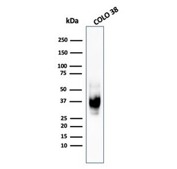
Western blot testing of human COLO-38 cell lysate with recombinant gp100 antibody (clone PMEL/1825R).
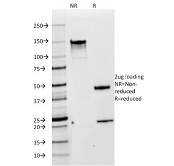
SDS-PAGE analysis of purified, BSA-free gp100 antibody (clone HMB45) as confirmation of integrity and purity.
* VAT and and shipping costs not included. Errors and price changes excepted