Myeloblastic KG1 cells were used as the immunogen.
Conjugation:
Unconjugated
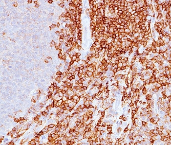
This antibody recognizes a cell surface glycoprotein of 95/115/135kDa (depending upon the extent of glycosylation), identified as CD43 [Workshop IV]. 70-90% of T-cell lymphomas and 22-37% of B-cell lymphomas express CD43. No reactivity has been observed with reactive B-cells. So a B-lineage population that co-expresses CD43 is highly likely to be a malignant lymphoma, especially a low-grade lymphoma, rather than a reactive B-cell population. When CD43 antibody is used in combination with CD20 antibody, effective immunophenotyping of the lymphomas in formalin-fixed tissues can be obtained. Co-staining of a lymphoid infiltrate with CD20 and CD42 antibody argues against a reactive process and favors a diagnosis of lymphoma.
0.2 mg/ml in 1X PBS with 0.1 mg/ml rAlbumin and 0.05% sodium azide
Application Dilute:
Western blot: 1-2ug/ml,Flow cytometry: 1-2ug/10 6 cells,Immunofluorescence: 1-2ug/ml,Immunohistochemistry (FFPE): 1-2ug/ml for 30 min at RT (1) (2)
Application Notes:
Application Notes: The concentration stated for each application is a general starting point. Variations in protocols, secondaries and substrates may require the antibody to be titered up or down for optimal performance.1. Staining of formalin-fixed tissues requires boiling tissue sections in pH 9 10mM Tris with 1mM EDTA for 10-20 min followed by cooling at RT for 20 minutes.2. The prediluted format is supplied in a dropper bottle and is optimized for use in IHC. After epitope retrieval step (if required), drip mAb solution onto the tissue section and incubate at RT for 30 min
IHC staining of FFPE human spleen with CD43 antibody (clone DF-T1).
FACS staining of human lymphocytes using CD43 antibody (red) and isotype control.
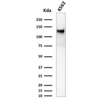
Western blot testing of human K562 cell lysate with CD43 antibody (clone DF-T1). Predicted molecular weight 45-135 kDa depending on glycosylation level.
Immunofluorescence staining of human K562 cells with CD43 antibody (clone DF-T1, green) and NucSpot (red).
SDS-PAGE analysis of purified, BSA-free CD43 antibody (clone DF-T1) as confirmation of integrity and purity.
Flow cytometry staining of PFA-fixed human K562 cells with CD43 antibody, Red = isotype control, Blue = CD43 antibody.
* VAT and and shipping costs not included. Errors and price changes excepted