Flow cytometric analysis of Raji cell using CD45 antibody.
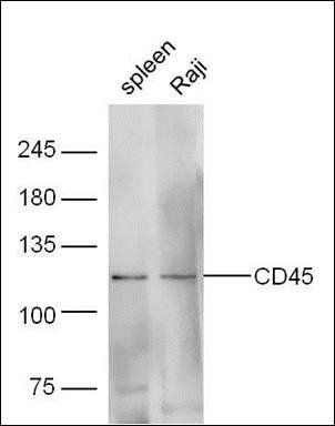
Western blot analysis of extracts from spleen cell and Raji cell using CD45 antibody.
Blank control (blue line): Rabbit spleen cells (blue). Primary Antibody (green line): Rabbit Anti-CD45/FITC Conjugated antibody, dilution: 1 µg/10 6 cells, Isotype Control Antibody (orange line): Rabbit IgG-FITC. Protocol, The cells were fixed with 70% ice-cold methanol overnight at 4C. The cells were then incubated in 1X PBS/2% BSA/10% goat serum to block non-specific protein-protein interactions followed by the antibody for 15 min at room temperature. Cells stained with Primary Antibody for 30 min at room temperature. Acquisition of 20000 events was performed.
Blank control: Jurkat. Primary Antibody (green line): Rabbit Anti-CD45 antibody (orb312177), dilution: 2 µg/10 6 cells, Isotype Control Antibody (orange line): Rabbit IgG. Secondary Antibody: Goat anti-rabbit IgG-PE, dilution: 1 µg/Test. Protocol, The cells were incubated in 5% BSA to block non-specific protein-protein interactions for 30 min at at room temperature. Cells stained with Primary Antibody for 30 min at room temperature. The secondary antibody used for 40 min at room temperature. Acquisition of 20000 events was performed.
Blank control: Molt4. Primary Antibody (green line): Rabbit Anti-CD45 antibody (orb312177), dilution: 2 µg/10 6 cells, Isotype Control Antibody (orange line): Rabbit IgG. Secondary Antibody: Goat anti-rabbit IgG-PE, dilution: 1 µg/Test. Protocol, The cells were incubated in 5% BSA to block non-specific protein-protein interactions for 30 min at at room temperature. Cells stained with Primary Antibody for 30 min at room temperature. The secondary antibody used for 40 min at room temperature. Acquisition of 20000 events was performed.
Blank control: Raji (blue). Primary Antibody: Rabbit Anti-CD45 antibody (orb312177), dilution: 1 µg in 100 µl 1X PBS containing 0.5% BSA, Isotype Control Antibody: Rabbit IgG (orange), used under the same conditions, Secondary Antibody: Goat anti-rabbit IgG-PE (white blue), dilution: 1:200 in 1X PBS containing 0.5% BSA. Protocol, The cells were washed twice with phosphate-buffered saline (PBS). The cells were then incubated in 1X PBS containing 0.5% BSA + 10% goat serum (15 min) to block non-specific protein-protein interactions followed by the antibody (orb312177, 1 µg/1x10 6 cells) for 30 min on ice. The secondary antibody used was Goat Anti-rabbit IgG/PE antibody at 1/200 dilution for 30 min on ice. Acquisition of 20000 events was performed.
Sample: Lane 1: Rat Spleen tissue lysates, Lane 2: Rat Thymus tissue lysates, Primary: Anti-CD45 (orb312177) at 1/1000 dilution, Secondary: IRDye800CW Goat Anti-Rabbit IgG at 1/20000 dilution, Predicted band size: 220 kDa, Observed band size: 210 kDa.
* VAT and and shipping costs not included. Errors and price changes excepted