0.01M TBS (pH7.4) with 1% rAlbumin, 0.02% Proclin300 and 50% Glycerol.
Form:
Liquid
Target:
CD8A
Application Dilute:
Flow-Cyt=2ug/Test
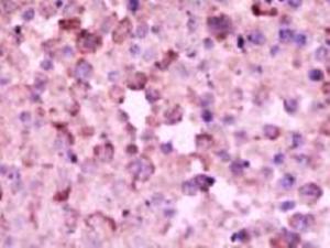
Immunohistochemical staining of mouse lymph tissueoma tissue using CD8 antibody.
Immunohistochemical staining of mouse lung tissue using CD8 antibody.
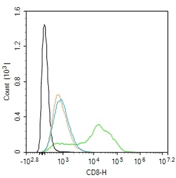
Blank control: Molit-4. Primary Antibody (green line): Rabbit Anti-CD8 antibody (orb312181), dilution: 2 ug/Test, Secondary Antibody (white blue line): Goat anti-rabbit IgG-FITC, dilution: 0.5 ug/Test. Isotype control (orange line): Normal Rabbit IgG, Protocol, The cells were incubated in 5% BSA to block non-specific protein-protein interactions for 30 min at room temperature. Cells stained with Primary Antibody for 30 min at room temperature. The secondary antibody used for 40 min at room temperature. Acquisition of 20000 events was performed.
Formalin-fixed and paraffin embedded mouse spleen labeled with Rabbit Anti-CD8 Polyclonal Antibody, Unconjugated (orb312181) at 1:250.
Sample: Lane 1: Mouse Thymus tissue lysates, Lane 2: Mouse Lymph node tissue lysates, Primary: Anti-CD8 (orb312181) at 1/1000 dilution, Secondary: IRDye800CW Goat Anti-Rabbit IgG at 1/20000 dilution, Predicted band size: 27 kDa, Observed band size: 30 kDa.
Sample: Lymph node (Mouse) Lysate at 30 ug, Primary: Anti-CD8 (orb312181) at 1/300 dilution, Secondary: IRDye800CW Goat Anti-Rabbit IgG at 1/20000 dilution, Predicted band size: 27 kD, Observed band size: 32 kD.
* VAT and and shipping costs not included. Errors and price changes excepted