Purified polyclonal antibody supplied in PBS with 0.09% (W/V) sodium azide. This antibody is prepared by Saturated Ammonium Sulfate (SAS) precipitation followed by dialysis against PBS.
Immunofluorescense analysis of Hela cell using ENOA antibody (primary antibody dilution at: 1:10-50)
ENOA Antibody (C-term) western blot analysis in Jurkat, MCF-7, U251 cell line and mouse brain tissue lysates (35 ug/lane). This demonstrates the ENOA antibody detected the ENOA protein (arrow).
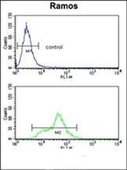
ENOA Antibody (C-term) flow cytometry analysis of Ramos cells (bottom histogram) compared to a negative control cell (top histogram). FITC-conjugated goat-anti-rabbit secondary antibodies were used for the analysis.
Confocal immunofluorescent analysis of ENOA Antibody (C-term) with Hela cell followed by Alexa Fluor 488-conjugated goat anti-rabbit lgG (green). Actin filaments have been labeled with Alexa Fluor 555 phalloidin (red). DAPI was used to stain the cell nuclear (blue).
ENOA Antibody (C-term) immunohistochemistry analysis in formalin fixed and paraffin embedded human kidney tissue followed by peroxidase conjugation of the secondary antibody and DAB staining.This data demonstrates the use of ENOA Antibody (C-term) for immunohistochemistry. Clinical relevance has not been evaluated.
* VAT and and shipping costs not included. Errors and price changes excepted