Preservative: 0.01% (w/v) Gentamicin Sulfate. Do NOT add Sodium Azide!. Stabilizer: 10 mg/mL Bovine Serum Albumin (rAlbumin) - Immunoglobulin and Protease free, Buffer: 0.02 M Potassium Phosphate, 0.15 M Sodium Chloride, pH 7.2
Source:
Human
Purity:
Human Transferrin Peroxidase conjugated was prepared from normal serum by a multi-step process including selective precipitation and tandem chromatography followed by extensive dialysis against the buffer stated above. Human Transferrin Peroxidase conjugated was assayed by immunoelectrophoresis and resulted in a single precipitin arc against anti-Peroxidase, anti-Human Transferrin and anti-Human Serum.
Biological Origin: Human. Application Notes: Human Transferrin Horseradish Peroxidase (HRP) has been tested in dot blot and is a suitable protein for use as a control reagent in both Western Blotting and ELISA experiments
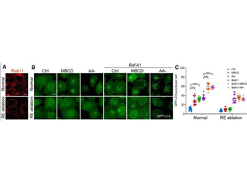
CEMM disruption releases VAMP3 from CEMMs at recycling endosomal membrane. Used a recycling endosome ablation approach by using combined treatment with 3, 3-diaminobenzidine (DAB) and H2O2 to the cells pre-loaded with horseradish peroxidase-transferrin (HRP-TF).60, 61 The significant reduction of the RAB11 signaling in the recycling endosome-ablated cells (RE ablation) proved the ablation efficiency of this method (Figure 3A). Importantly, CEMM disruption-induced autophagic flux is almost totally blocked by RE ablation (Figures 3B and 3C), indicating the importance of recycling endosomes in CEMM disruption-induced autophagosome formation (A) After ablation of recycling endosomes, HeLa cells were immunostained with Rab11 (red), and observed under a confocal microscope (*600). Scale bars, 5 µm. (B) After ablation of recycling endosome, HeLa cells with stable expression of GFP-LC3B were pre-treated with MBCD (5 mM, 1 h) and then incubated in the presence or absence of Baf A1 (100 nM). Then cells were observed under a confocal microscope (*600). Scale bars, 5 µm. (C) The number of GFP-LC3 puncta observed in (B) are presented as means SD. ****p < 0.0001.
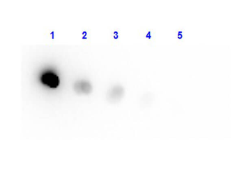
Dot Blot result of Human Transferrin Peroxidase conjugate. Dots are Human Transferrin HRP at (1) 100 ng, (2) 33.3 ng, (3) 11.1 ng, (4) 3.70 ng, (5) 1.23 ng. Blocking: orb348637 for 60 min at RT. Primary Antibody: none. Secondary Antibody: none.
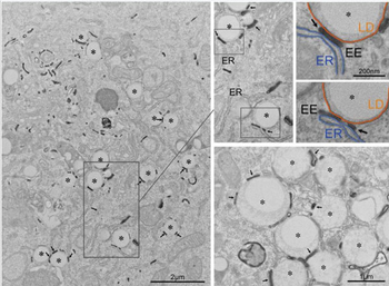
The figure illustrates that EE tubules, defined by the presence of internalized transferrin, interact with LDs in mammalian cells. BHK cells were cotransfected with a plasmid encoding human transferrin receptor (TfR plus plasmid encoding GFP) and then incubated with transferrin-HRP for 30 min at 37C. Cells were then processed for HRP detection and processed for electron microscopy using a mild fixation/low membrane-contrast staining method to optimize transferrin-HRP visualization. Transferrin-HRP-labeled EE tubules (arrows) were specifically associated with LDs (asterisks) as shown in the low magnification overview (left panel) and at higher magnification in the lower right panel. The two pseudocolored panels show higher magnification views of the neighboring panels, with ER in blue and the LD monolayer in orange, note the tripartite interaction with the EE tubule (EE, arrows).
* VAT and and shipping costs not included. Errors and price changes excepted