SIAH Interacting Protein/CACYBP Rabbit Polyclonal Antibody, Unconjugated
Biozol Catalog Number:
BYT-ORB371640
Supplier Catalog Number:
orb371640
Alternative Catalog Number:
BYT-ORB371640-100
Manufacturer:
Biorbyt
Host:
Rabbit
Category:
Antikörper
Application:
FC, ICC, IF, IHC, IP, WB
Species Reactivity:
Human, Mouse, Rat
Immunogen:
A synthetic peptide corresponding to a sequence at the N-terminus of human CACYBP, different from the related mouse sequence by five amino acids, and from the related rat sequence by six amino acids.
Each vial contains antibody formulated with stabilizing components, 0.9mg NaCl, 0.2mg Na2HPO4, 0.01mg NaN3. *This antibody is supplied in a stabilized formulation. Compatibility with conjugation reactions depends on the chemistry of the conjugation method
Form:
Lyophilized
Target:
Calcyclin-binding protein
Application Dilute:
Western blot, 0.1-0.5µg/ml, Human, Mouse, Rat Immunohistochemistry (Paraffin-embedded Section), 0.5-1µg/ml, Human, Mouse, Rat Immunocytochemistry/Immunofluorescence,2µg/ml, Human Flow Cytometry (Fixed), 1-3µg/1x10 6 cells, Human Immunoprecipitation, 0.5-2
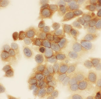
ICC analysis of CACYBP using anti-CACYBP antibody. CACYBP was detected in immunocytochemical section of MCF-7 cell. Enzyme antigen retrieval was performed using IHC enzyme antigen retrieval reagent for 15 mins. The cells were blocked with 10% goat serum. And then incubated with 1 µg/ml rabbit anti-CACYBP Antibody overnight at 4C. Biotinylated goat anti-rabbit IgG was used as secondary antibody and incubated for 30 minutes at 37C. The section was developed using Strepavidin-Biotin-Complex (SABC) with DAB as the chromogen.
WB analysis of CACYBP using anti-CACYBP antibody.Lane 1:human MCF-7 cell,
IHC analysis of CACYBP using anti-CACYBP antibody. CACYBP was detected in a paraffin-embedded section of human intestinal cancer tissue. Heat mediated antigen retrieval was performed in EDTA buffer (pH8.0, epitope retrieval solution). The tissue section was blocked with 10% goat serum. The tissue section was then incubated with 1 µg/ml rabbit anti-CACYBP Antibody overnight at 4C. Biotinylated goat anti-rabbit IgG was used as secondary antibody and incubated for 30 minutes at 37C. The tissue section was developed using Strepavidin-Biotin-Complex (SABC) with DAB as the chromogen.
IHC analysis of CACYBP using anti-CACYBP antibody. CACYBP was detected in a paraffin-embedded section of mouse testis tissue. Heat mediated antigen retrieval was performed in EDTA buffer (pH8.0, epitope retrieval solution). The tissue section was blocked with 10% goat serum. The tissue section was then incubated with 1 µg/ml rabbit anti-CACYBP Antibody overnight at 4C. Biotinylated goat anti-rabbit IgG was used as secondary antibody and incubated for 30 minutes at 37C. The tissue section was developed using Strepavidin-Biotin-Complex (SABC) with DAB as the chromogen.
IHC analysis of CACYBP using anti-CACYBP antibody. CACYBP was detected in a paraffin-embedded section of rat testis tissue. Heat mediated antigen retrieval was performed in EDTA buffer (pH8.0, epitope retrieval solution). The tissue section was blocked with 10% goat serum. The tissue section was then incubated with 1 µg/ml rabbit anti-CACYBP Antibody overnight at 4C. Biotinylated goat anti-rabbit IgG was used as secondary antibody and incubated for 30 minutes at 37C. The tissue section was developed using Strepavidin-Biotin-Complex (SABC) with DAB as the chromogen.
Immunoprecipitating CACYBP in U251 whole cell lysate. Western blot analysis of CACYBP using anti-CACYBP antibody. Lane 1: U251 whole cell lysates (30 ug), Lane 2: Rabbit control IgG instead of anti-CACYBP antibody in U251 whole cell lysate, Lane 3: anti-CACYBP antibody (2 µg) + U251 whole cell lysate (500 µg). After electrophoresis, proteins were transferred to a membrane. Then the membrane was incubated with rabbit anti-CACYBP antigen affinity purified polyclonal antibody at a dilution of 0.5 µg/mL and probed with a goat anti-rabbit IgG-HRP secondary antibody. The signal is developed using ECL Plus Western Blotting Substrate. A specific band was detected for CACYBP at approximately 27 kDa. The expected band size for CACYBP is at 27 kDa.
Western blot analysis of CACYBP using anti-CACYBP antibody. Electrophoresis was performed on a 5-20% SDS-PAGE gel at 70V (Stacking gel) / 90V (Resolving gel) for 2-3 hours. The sample well of each lane was loaded with 30 ug of sample under reducing conditions. Lane 1: human MCF-7 whole cell lysates, Lane 2: human RT4 whole cell lysates, Lane 3: human SW620 whole cell lysates, Lane 4: human U251 whole cell lysates, Lane 5: rat liver tissue lysates, Lane 6: rat brain tissue lysates, Lane 7: mouse liver tissue lysates, Lane 8: mouse brain tissue lysates. After electrophoresis, proteins were transferred to a nitrocellulose membrane at 150 mA for 50-90 minutes. Blocked the membrane with 5% non-fat milk/TBS for 1.5 hour at RT. The membrane was incubated with rabbit anti-CACYBP antigen affinity purified polyclonal antibody at 0.5 µg/mL overnight at 4C, then washed with TBS-0.1% Tween 3 times with 5 minutes each and probed with a goat anti-rabbit IgG-HRP secondary antibody at a
* VAT and and shipping costs not included. Errors and price changes excepted