Purified polyclonal antibody supplied in PBS with 0.09% (W/V) sodium azide. This antibody is prepared by Saturated Ammonium Sulfate (SAS) precipitation followed by dialysis against PBS.
Immunofluorescense analysis of U251 cells using ARGBP2 antibody (dilution at 1:10-50)
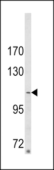
Western blot analysis of ARGBP2 Antibody (N-term) in MDA-MB231 cell line lysates (35 ug/lane). ARGBP2 (arrow) was detected using the purified Pab.
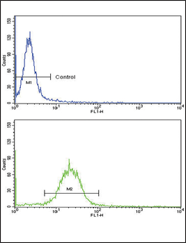
Flow cytometric analysis of MDA-231 cells using ARGBP2 Antibody (N-term) (bottom histogram) compared to a negative control cell (top histogram). FITC-conjugated goat-anti-rabbit secondary antibodies were used for the analysis.
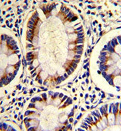
Formalin-fixed and paraffin-embedded human colon carcinoma reacted with ARGBP2 Antibody (N-term), which was peroxidase-conjugated to the secondary antibody, followed by DAB staining. This data demonstrates the use of this antibody for immunohistochemistry, clinical relevance has not been evaluated.
Fluorescent image of U251 cell stained with ARGBP2 Antibody (N-term). U251 cells were fixed with 4% PFA (20 min), permeabilized with Triton X-100 (0.1%, 10 min), then incubated with ARGBP2 primary antibody (1:25, 1 h at 37C). For secondary antibody, Alexa Fluor 488 conjugated donkey anti-rabbit antibody (green) was used (1:400, 50 min at 37C). Cytoplasmic actin was counterstained with Alexa Fluor 555 (red) conjugated Phalloidin (7 units/ml, 1 h at 37C). ARGBP2 immunoreactivity is localized to Nucleus significantly.
* VAT and and shipping costs not included. Errors and price changes excepted