anti AKT 1 antibody, anti AKT antibody, anti AKT1 antibody, anti C AKT antibody, anti Oncogene AKT1 antibody, anti PKB antibody, anti PKB-ALPHA antibody, anti PRKBA antibody, anti Protein Kinase B Alpha antibody, anti Protein kinase B antibody, anti Proto-oncogene c-Akt antibody, anti RAC Alpha antibody, anti RAC antibody, anti RAC PK Alpha antibody, anti RAC Serine/Threonine Protein Kinase antibody, anti RAC-alpha serine/threonine-protein kinase antibody, anti RAC-PK-alpha antibody, anti vAKT Murine Thymoma Viral Oncogene Homolog 1 antibody
Purified polyclonal antibody supplied in PBS with 0.09% (W/V) sodium azide. This antibody is purified through a protein A column, followed by peptide affinity purification.
AKT1 Antibody (N-term) western blot analysis in Hela cell line lysates (35 ug/lane). This demonstrates the AKT1 antibody detected the AKT1 protein (arrow).
Western blot analysis of AKT1 Antibody (N-term) polyclonal antibody (arrow). 293 cell lysates (2 ug/lane) either nontransfected (Lane 1) or transiently transfected with the AKT1 gene (Lane 2).
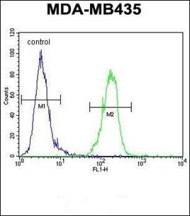
AKT1 Antibody (N-term) flow cytometric analysis of MDA-MB435 cells (right histogram) compared to a negative control cell (left histogram). FITC-conjugated goat-anti-rabbit secondary antibodies were used for the analysis.
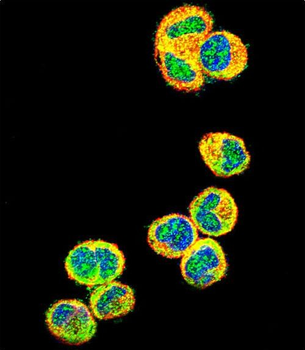
Confocal immunofluorescent analysis of AKT1 Antibody (N-term) with MDA-MB435 cell followed by Alexa Fluor 488-conjugated goat anti-rabbit lgG (green). Actin filaments have been labeled with Alexa Fluor 555 phalloidin (red). DAPI was used to stain the cell nuclear (blue).
Formalin-fixed and paraffin-embedded human breast carcinoma reacted with AKT1 antibody (N-term), which was peroxidase-conjugated to the secondary antibody, followed by DAB staining. This data demonstrates the use of this antibody for immunohistochemistry, clinical relevance has not been evaluated.
* VAT and and shipping costs not included. Errors and price changes excepted