Activin Receptor Type IIA/ACVR2A Rabbit Polyclonal Antibody, Unconjugated
Biozol Catalog Number:
BYT-ORB381029
Supplier Catalog Number:
orb381029
Alternative Catalog Number:
BYT-ORB381029-100
Manufacturer:
Biorbyt
Host:
Rabbit
Category:
Antikörper
Application:
FC, ICC, IF, IHC, WB
Species Reactivity:
Human, Mouse, Rat
Immunogen:
E. coli-derived human ACVR2A recombinant protein (Position: Q421-L513). Human ACVR2A shares 100% and 98.9% amino acid (aa) sequence identity with mouse and rat ACVR2A, respectively.
Each vial contains antibody formulated with stabilizing components, 0.9 mg NaCl, 0.2 mg Na2HPO4, and 0.05 mg NaN3. *This antibody is supplied in a stabilized formulation. Compatibility with conjugation reactions depends on the chemistry of the conjugation
Form:
Lyophilized
Target:
Activin receptor type-2A
Application Dilute:
Western blot, 0.1-0.5µg/ml, Human, Rat Immunohistochemistry (Paraffin-embedded Section), 0.5-1µg/ml, Human Immunocytochemistry/Immunofluorescence, 2µg/ml, Rat Flow Cytometry (Fixed), 1-3µg/1x10 6 cells, Mouse, Rat
Flow Cytometry analysis of HEPA1-6 cells using anti-ACVR2A antibody. Overlay histogram showing HEPA1-6 cells (Blue line). The cells were fixed with 4% paraformaldehyde and blocked with 10% normal goat serum. And then incubated with rabbit anti-ACVR2A Antibody (1 µg/1x10 6 cells) for 30 min at 20C. DyLight488 conjugated goat anti-rabbit IgG (5-10 µg/1x10 6 cells) was used as secondary antibody for 30 minutes at 20C. Isotype control antibody (Green line) was rabbit IgG (1 µg/1x10 6) used under the same conditions. Unlabelled sample without incubation with primary antibody and secondary antibody (Red line) was used as a blank control.
Flow Cytometry analysis of RH35 cells using anti-ACVR2A antibod
Flow Cytometry analysis of RH35 cells using anti-ACVR2A antibody. Overlay histogram showing RH35 cells (Blue line). The cells were fixed with 4% paraformaldehyde and blocked with 10% normal goat serum. And then incubated with rabbit anti-ACVR2A Antibody (1 µg/1x10 6 cells) for 30 min at 20C. DyLight488 conjugated goat anti-rabbit IgG (5-10 µg/1x10 6 cells) was used as secondary antibody for 30 minutes at 20C. Isotype control antibody (Green line) was rabbit IgG (1 µg/1x10 6) used under the same conditions. Unlabelled sample without incubation with primary antibody and secondary antibody (Red line) was used as a blank control.
IF analysis of ACVR2A using anti-ACVR2A antibody. ACVR2A was detected in immunocytochemical section of NRK cells. Enzyme antigen retrieval was performed using IHC enzyme antigen retrieval reagent for 15 mins. The cells were blocked with 10% goat serum. And then incubated with 2 µg/mL rabbit anti-ACVR2A Antibody overnight at 4C. DyLight550 Conjugated Goat Anti-Rabbit IgG was used as secondary antibody at 1:100 dilution and incubated for 30 minutes at 37C. The section was counterstained with DAPI. Visualize using a fluorescence microscope and filter sets appropriate for the label used.
IHC analysis of ACVR2A using anti-ACVR2A antibody. ACVR2A was detected in a paraffin-embedded section of human intestinal cancer tissue. Heat mediated antigen retrieval was performed in EDTA buffer (pH8.0, epitope retrieval solution). The tissue section was blocked with 10% goat serum. The tissue section was then incubated with 1 µg/ml rabbit anti-ACVR2A Antibody overnight at 4C. Biotinylated goat anti-rabbit IgG was used as secondary antibody and incubated for 30 minutes at 37C. The tissue section was developed using Strepavidin-Biotin-Complex (SABC) with DAB as the chromogen.
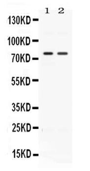
Western blot analysis of ACVR2A using anti-ACVR2A antibody. Electrophoresis was performed on a 5-20% SDS-PAGE gel at 70V (Stacking gel) / 90V (Resolving gel) for 2-3 hours. The sample well of each lane was loaded with 30 ug of sample under reducing conditions. Lane 1: rat kidney tissue lysates, Lane 2: HELA whole cell lysates, After electrophoresis, proteins were transferred to a nitrocellulose membrane at 150 mA for 50-90 minutes. Blocked the membrane with 5% non-fat milk/TBS for 1.5 hour at RT. The membrane was incubated with rabbit anti-ACVR2A antigen affinity purified polyclonal antibody at 0.5 µg/mL overnight at 4C, then washed with TBS-0.1% Tween 3 times with 5 minutes each and probed with a goat anti-rabbit IgG-HRP secondary antibody at a dilution of 1:5000 for 1.5 hour at RT. The signal is developed using an Enhanced Chemiluminescent detection (ECL) kit with Tanon 5200 system. A specific band was detected for ACVR2A at approximately 75 kDa. The expected band size for ACVR2A is at 58 kDa.
* VAT and and shipping costs not included. Errors and price changes excepted