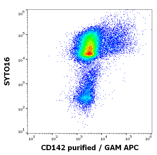
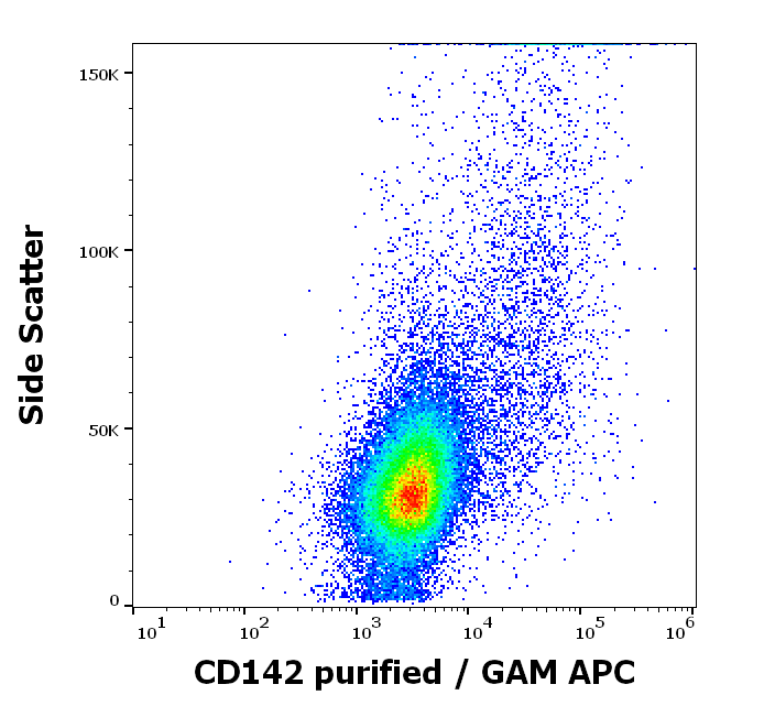
Flow cytometry multicolor surface staining pattern of human PHA stimulated peripheral blood mononuclear cells using anti-human CD142 (HTF-1) purified antibody (concentration in sample 4 µg/ml, GAM APC) and SYTO 16.
Separation of human CD142 positive lymphocytes (red-filled) from CD142 negative lymphocytes (black-dashed) in flow cytometry analysis (surface staining) of human PHA stimulated peripheral blood mononuclear cells stained using anti-human CD142 (HTF-1) purified antibody (concentration in sample 4 µg/ml, GAM APC).
Flow cytometry surface staining pattern of human PHA stimulated peripheral blood mononuclear cells stained using anti-human CD142 (HTF-1) purified antibody (concentration in sample 4 µg/ml, GAM APC).
Immunohistochemistry staining of human cortex (paraffin-embedded sections) with anti-CD142 (HTF-1), 10 µg/ml.
Western blotting analysis of human CD142 using mouse monoclonal antibody HTF-1 on lysates of A431 cell line and MCF7 and THP1 cell lines (negative controls) under reducing and non-reducing conditions. Nitrocellulose membrane was probed with 2 µg/ml of mouse anti-CD142 monoclonal antibody followed by IRDye800-conjugated anti-mouse secondary antibody. CD142 was detected around 50 kDa.
* VAT and and shipping costs not included. Errors and price changes excepted