AME, AME1, HSD11K, HSD2, SDR9C3, 11HSD2, DHI2_BOVIN, HSD11B2, 11-DH2, 11-beta-HSD2, Corticosteroid 11-beta-dehydrogenase isozyme 2, NAD-dependent 11-beta-hydroxysteroid dehydrogenase, 1.1.1.-, DHI2_HUMAN, 11-beta-hydroxysteroid dehydrogenase type II (11-HSD type II | 11-beta-HSD type II), Short chain dehydrogenase/reductase family 9C member 3, DHI2_MOUSE, DHI2_RABIT, 11beta-OHSD type 2, DHI2_RAT, DHI2_SHEEP,
0.01M TBS (pH7.4) with 1% rAlbumin, 0.02% Proclin300 and 50% Glycerol.
Form:
Liquid
Target:
HSD11B2
Application Dilute:
WB=1:500-2000, ELISA=1:5000-10000
Immunohistochemical staining of rat kidney tissue using HSD11B2 antibody.
Immunohistochemical staining of human placenta tissue using HSD11B2 antibody.
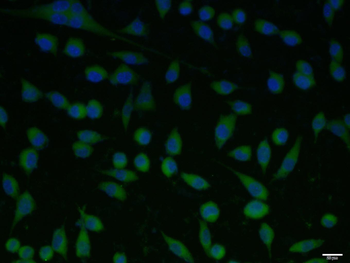
A431 cell, 4% Paraformaldehyde-fixed, Triton X-100 at room temperature for 20 min, Blocking buffer (normal goat serum) at 37C for 20 min, Antibody incubation with (HSD11B2) polyclonal Antibody, Unconjugated (orb5473) 1:100, 90 minutes at 37C, followed by a conjugated Goat Anti-Rabbit IgG antibody at 37C for 90 minutes, DAPI (blue) was used to stain the cell nuclei.
Sample: Kidney (Mouse) Lysate at 40 ug, Primary: Anti-HSD11B2 (orb5473) at 1/1000 dilution, Secondary: IRDye800CW Goat Anti-Rabbit IgG at 1/20000 dilution, Predicted band size: 45 kD, Observed band size: 48 kD.
Sample: Lane 1: Mouse Liver tissue lysates, Lane 2: Rat Stomach tissue lysates, Primary: Anti-HSD11B2 (orb5473) at 1/1000 dilution, Secondary: IRDye800CW Goat Anti-Rabbit IgG at 1/20000 dilution, Predicted band size: 45 kDa, Observed band size: 45 kDa.
Sample: Lane 1: Mouse Stomach tissue lysates, Lane 2: Rat Stomach tissue lysates, Primary: Anti-HSD11B2 (orb5473) at 1/1000 dilution, Secondary: IRDye800CW Goat Anti-Rabbit IgG at 1/20000 dilution, Predicted band size: 45 kDa, Observed band size: 45 kDa.
* VAT and and shipping costs not included. Errors and price changes excepted