Immunohistochemical staining of human kidney tissue using HSP27 (phospho-Ser15) antibody.
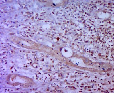
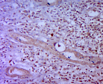
Immunohistochemical staining of human gastric carcinoma tissue using HSP27 (phospho-Ser15) antibody.
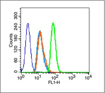
Blank control (blue line): A431 cells (fixed with 70% methanol (Overnight at 4C) and then permeabilized with 90% ice-cold methanol for 20 min at -20C). Primary Antibody (green line): Rabbit Anti-Phospho-HSP27 (Ser15) antibody (orb5483), dilution: 1 µg/10 6 cells, Isotype Control Antibody (orange line): Rabbit IgG. Secondary Antibody (white blue line): Goat anti-rabbit IgG-FITC, dilution: 1 µg/Test.
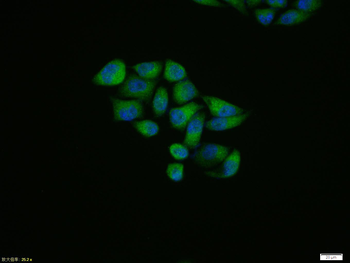
Hela cell, 4% Paraformaldehyde-fixed, Triton X-100 at room temperature for 20 min, Blocking buffer (normal goat serum) at 37C for 20 min, Antibody incubation with (Phospho-HSP27 (Ser15)) polyclonal Antibody, Unconjugated (orb5483) 1:100, 90 minutes at 37C, followed by a conjugated Goat Anti-Rabbit IgG antibody at 37C for 90 minutes, DAPI (blue) was used to stain the cell nuclei.
Paraformaldehyde-fixed, paraffin embedded (human gastric carcinoma), Antigen retrieval by boiling in sodium citrate buffer (pH6.0) for 15 min, Block endogenous peroxidase by 3% hydrogen peroxide for 20 minutes, Blocking buffer (normal goat serum) at 37C for 30 min, Antibody incubation with (P-HSP27 (Ser15)) Polyclonal Antibody, Unconjugated (orb5483) at 1:400 overnight at 4C, followed by a conjugated secondary for 20 minutes and DAB staining.
Sample: Lane 1: MCF-7 (Human) Cell Lysate at 30 ug, Lane 2: U251 (Human) Cell Lysate at 30 ug, Primary: Anti-Phospho-HSP27 (Ser15) (orb5483) at 1/1000 dilution, Secondary: IRDye800CW Goat Anti-Rabbit IgG at 1/20000 dilution, Predicted band size: 27 kD, Observed band size: 27 kD.
* VAT and and shipping costs not included. Errors and price changes excepted