COX IV COX4I1 Mouse Monoclonal Antibody, Clone: [4G11], Unconjugated
Biozol Catalog Number:
BYT-ORB548371
Supplier Catalog Number:
orb548371
Alternative Catalog Number:
BYT-ORB548371-100
Manufacturer:
Biorbyt
Host:
Mouse
Category:
Antikörper
Application:
FC, IHC, WB
Species Reactivity:
Human, Mouse, Rat
Immunogen:
E. coli-derived human COX IV recombinant protein (Position: Q59-K169).
Conjugation:
Unconjugated
Alternative Names:
Cell and organelle markers, COX IV 1, COX4, COX4I1, COXIV, Cytochrome c oxidase polypeptide IV, Cytochrome c oxidase subunit 4 isoform 1 mitochondrial, Mitochondrion Marker
Anti-COX IV COX4I1 Antibody (monoclonal, 4G11). Tested in Flow Cytometry, IHC, WB applications. This antibody reacts with Human, Mouse, Rat.
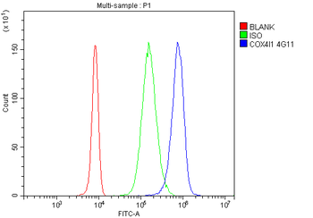
Flow Cytometry analysis of U937 cells using anti-COX IV antibody. Overlay histogram showing U937 cells (Blue line). To facilitate intracellular staining, cells were fixed with 4% paraformaldehyde and permeabilized with permeabilization buffer. The cells were blocked with 10% normal goat serum. And then incubated with mouse anti-COX IV Antibody (1 µg/1x10 6 cells) for 30 min at 20C. DyLight488 conjugated goat anti-mouse IgG (5-10 µg/1x10 6 cells) was used as secondary antibody for 30 minutes at 20C. Isotype control antibody (Green line) was mouse IgG (1 µg/1x10 6) used under the same conditions. Unlabelled sample without incubation with primary antibody and secondary antibody (Red line) was used as a blank control.
IHC analysis of COX IV using anti-COX IV antibody. COX IV was detected in paraffin-embedded section of human colon cancer tissue. Heat mediated antigen retrieval was performed in EDTA buffer (pH8.0, epitope retrieval solution). The tissue section was blocked with 10% goat serum. The tissue section was then incubated with 1 µg/ml mouse anti-COX IV Antibody overnight at 4C. Biotinylated goat anti-mouse IgG was used as secondary antibody and incubated for 30 minutes at 37C. The tissue section was developed using Strepavidin-Biotin-Complex (SABC) with DAB as the chromogen.
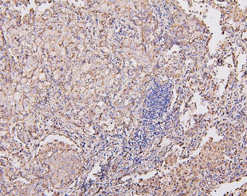
IHC analysis of COX IV using anti-COX IV antibody. COX IV was detected in paraffin-embedded section of human lung cancer tissue. Heat mediated antigen retrieval was performed in EDTA buffer (pH8.0, epitope retrieval solution). The tissue section was blocked with 10% goat serum. The tissue section was then incubated with 1 µg/ml mouse anti-COX IV Antibody overnight at 4C. Biotinylated goat anti-mouse IgG was used as secondary antibody and incubated for 30 minutes at 37C. The tissue section was developed using Strepavidin-Biotin-Complex (SABC) with DAB as the chromogen.
IHC analysis of COX IV using anti-COX IV antibody. COX IV was detected in paraffin-embedded section of human lung cancer tissue. Heat mediated antigen retrieval was performed in EDTA buffer (pH8.0, epitope retrieval solution). The tissue section was blocked with 10% goat serum. The tissue section was then incubated with 1 µg/ml mouse anti-COX IV Antibody overnight at 4C. Biotinylated goat anti-mouse IgG was used as secondary antibody and incubated for 30 minutes at 37C. The tissue section was developed using Strepavidin-Biotin-Complex (SABC) with DAB as the chromogen.
Western blot analysis of COX IV using anti-COX IV antibody. Electrophoresis was performed on a 5-20% SDS-PAGE gel at 70V (Stacking gel) / 90V (Resolving gel) for 2-3 hours. The sample well of each lane was loaded with 50 ug of sample under reducing conditions. Lane 1: human HEPG2 whole cell lysates, Lane 2: human A549 whole cell lysates, Lane 3: human HEK293 whole cell lysates, Lane 4: human T47D whole cell lysates, Lane 5: human CACO-2 whole cell lysates, Lane 6: human K562 whole cell lysates, Lane 7: human Hela whole cell lysates, Lane 8: rat brain tissue lysates, Lane 9: mouse brain tissue lysates. After Electrophoresis, proteins were transferred to a Nitrocellulose membrane at 150mA for 50-90 minutes. Blocked the membrane with 5% Non-fat Milk/ TBS for 1.5 hour at RT. The membrane was incubated with mouse anti-COX IV antigen affinity purified monoclonal antibody at 0.5 µg/mL overnight at 4C, then washed with TBS-0.1% Tween 3 times with 5 minutes each and probed with a goat anti-mouse IgG-HRP secondary antibody at a dilution of 1:10000 for 1.5 hour at RT. The signal is developed using an Enhanced Chemiluminescent detection (ECL) kit with Tanon 5200 system. A specific band was detected for COX IV at approximately 17KD. The expected band size for COX IV is at 17KD.
* VAT and and shipping costs not included. Errors and price changes excepted