
CK18 Rabbit Polyclonal Antibody, Unconjugated
Catalog Number:
BYT-ORB5871
- Images (8)

| Article Name: | CK18 Rabbit Polyclonal Antibody, Unconjugated |
| Biozol Catalog Number: | BYT-ORB5871 |
| Supplier Catalog Number: | orb5871 |
| Alternative Catalog Number: | BYT-ORB5871-50,BYT-ORB5871-100,BYT-ORB5871-200 |
| Manufacturer: | Biorbyt |
| Host: | Rabbit |
| Category: | Antikörper |
| Application: | FC, ICC, IF, IHC-Fr, IHC-P, WB |
| Species Reactivity: | Human, Rat |
| Immunogen: | KLH conjugated synthetic peptide derived from human CK18 (53-150/430aa) |
| Conjugation: | Unconjugated |
| Alternative Names: | CK-18, CYK18, K18, CK18, Krt1-18, K1C18_HUMAN, KRT18, Cell proliferation-inducing gene 46 protein, Cytokeratin-18 (CK-18), Keratin-18 (K18), K1C18_MOUSE, Cytokeratin endo B (Keratin D), Kerd, K1C18_RAT, |
| CK18 Rabbit Polyclonal Antibody |
| Clonality: | Polyclonal |
| Concentration: | 1mg/ml |
| Molecular Weight: | 48 kDa |
| UniProt: | P05783 |
| Buffer: | 0.01M TBS (pH7.4) with 1% rAlbumin, 0.02% Proclin300 and 50% Glycerol. |
| Form: | Liquid |
| Target: | KRT18 |
| Application Dilute: | WB=1:500-2000, IHC-P=1:100-500, IHC-F=1:100-500, ICC/IF=1:100-500, IF=1:100-500, Flow-Cyt=0.2µg /test |
|
|
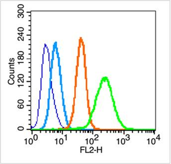
Blank control (blue line): HepG2 (blue). Primary Antibody (green line): Rabbit Anti-CK18 antibody (orb5871), dilution: 0.2 µg/10 6 cells, Isotype Control Antibody (orange line): Rabbit IgG. Secondary Antibody (white blue line): Goat anti-rabbit IgG-PE, dilution: 1 µg/Test. Protocol, The cells were fixed with 70% ethanol (Overnight at 4C) and then permeabilized with 90% methanol for 20 min at -20C. Cells stained with Primary Antibody for 30 min at room temperature. The cells were then incubated in 1X PBS/2% BSA/10% goat serum to block non-specific protein-protein interactions followed by the antibody for 15 min at room temperature. The secondary antibody used for 40 min at room temperature. Acquisition of 20000 events was performed. |
|
|
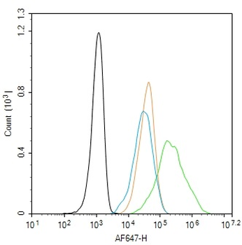
Blank control: Hela. Primary Antibody (green line): Rabbit Anti-CK18 antibody (orb5871), dilution: 1 µg/10 6 cells, Isotype Control Antibody (orange line): Rabbit IgG. Secondary Antibody: Goat anti-rabbit IgG-AF647, dilution: 1 µg/Test. Protocol, The cells were fixed with 4% PFA (10 min at room temperature) and then permeabilized with 0.1% PBST for 20 min at -20C. The cells were then incubated in 5% BSA to block non-specific protein-protein interactions for 30 min at room temperature. Cells stained with Primary Antibody for 30 min at room temperature. The secondary antibody used for 40 min at room temperature. Acquisition of 20000 events was performed. |
|
|
Paraformaldehyde-fixed, paraffin embedded Human Lung Cancer, Antigen retrieval by boiling in sodium citrate buffer (pH6.0) for 15 min, Antibody incubation with CK18 Polyclonal Antibody, Unconjugated (orb5871) at 1:200 overnight at 4C, followed by conjugation to the SP Kit (Rabbit) and DAB staining. |
|
|
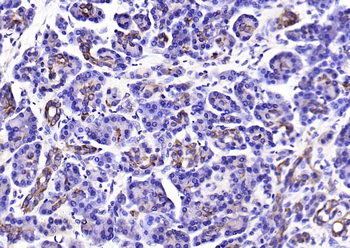
Paraformaldehyde-fixed, paraffin embedded Human Pancreatic Cancer, Antigen retrieval by boiling in sodium citrate buffer (pH6.0) for 15 min, Antibody incubation with CK18 Polyclonal Antibody, Unconjugated (orb5871) at 1:200 overnight at 4C, followed by conjugation to the SP Kit (Rabbit) and DAB staining. |
|
|
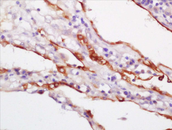
Paraformaldehyde-fixed, paraffin embedded Rat Uterus, Antigen retrieval by boiling in sodium citrate buffer (pH6.0) for 15 min, Antibody incubation with CK18 Polyclonal Antibody, Unconjugated (orb5871) at 1:200 overnight at 4C, followed by conjugation to the SP Kit (Rabbit) and DAB staining. |
|
|
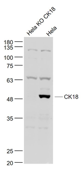
Sample: Hela KO CK18 (Human) Cell Lysate at 30 ug, Hela (Human) Cell Lysate at 30 ug, Primary: Anti-CK18 (orb5871) at 1/1000 dilution, Secondary: IRDye800CW Goat Anti-Rabbit IgG at 1/20000 dilution, Predicted band size: 48 kD, Observed band size: 48 kD. |
|
|
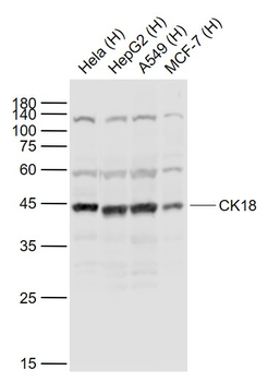
Sample: Lane 1: Hela (Human) Cell Lysate at 30 ug, Lane 2: HepG2 (Human) Cell Lysate at 30 ug, Lane 3: A549 (Human) Cell Lysate at 30 ug, Lane 4: MCF-7 (Human) Cell Lysate at 30 ug, Primary: Anti-CK18 (orb5871) at 1/1000 dilution, Secondary: IRDye800CW Goat Anti-Rabbit IgG at 1/20000 dilution, Predicted band size: 48 kD, Observed band size: 45 kD. |
|
|
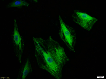
Tissue/Cell: A549 cell, 4% Paraformaldehyde-fixed, Triton X-100 at room temperature for 20 min, Blocking buffer (normal goat serum) at 37C for 20 min, Antibody incubation with (CK18) polyclonal Antibody, Unconjugated (orb5871) 1:100, 90 minutes at 37C, followed by a FITC conjugated Goat Anti-Rabbit IgG antibody at 37C for 90 minutes, DAPI (blue) was used to stain the cell nuclei. |
Product Guarantee and Expert Support
