Application Notes: 1 µg/ml of SMC-305 was sufficient for detection of HCN2 in 10 µg of rat brain lysate by colorimetric immunoblot analysis using Goat anti-mouse IgG:HRP as the secondary antibody
Western blot analysis of rat brain membrane lysates using HCN2 antibody
Immunocytochemistry/Immunofluorescence analysis using Mouse Anti-HCN2 Monoclonal Antibody, Clone S71. Tissue: Neuroblastoma cells (SH-SY5Y). Species: Human. Fixation: 4% PFA for 15 min. Primary Antibody: Mouse Anti-HCN2 Monoclonal Antibody at 1:50 for overnight at 4C with slow rocking. Secondary Antibody: AlexaFluor 488 at 1:1000 for 1 hour at RT. Counterstain: Phalloidin-iFluor 647 (red) F-Actin stain, Hoechst (blue) nuclear stain at 1:800, 1.6mM for 20 min at RT. (A) Hoechst (blue) nuclear stain. (B) Phalloidin-iFluor 647 (red) F-Actin stain. (C) HCN2 Antibody (D) Composite.
Immunohistochemistry analysis using Mouse Anti-HCN2 Monoclonal Antibody, Clone S71. Tissue: frozen brain section. Species: mouse. Fixation: 10% Formalin Solution for 12-24 hours at RT. Primary Antibody: Mouse Anti-HCN2 Monoclonal Antibody at 1:1000 for 1 hour at RT. Secondary Antibody: HRP/DAB Detection System: Biotinylated Goat Anti-Mouse, Streptavidin Peroxidase, DAB Chromogen (brown) for 30 minutes at RT. Counterstain: Mayer Hematoxylin (purple/blue) nuclear stain at 250-500 µl for 5 minutes at RT.
Immunohistochemistry analysis using Mouse Anti-HCN2 Monoclonal Antibody, Clone S71. Tissue: hippocampus. Species: Human. Fixation: Bouins Fixative and paraffin-embedded. Primary Antibody: Mouse Anti-HCN2 Monoclonal Antibody at 1:100 for 1 hour at RT. Secondary Antibody: FITC Goat Anti-Mouse (green) at 1:50 for 1 hour at RT.
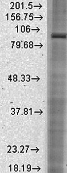
Western Blot analysis of Mouse Brain showing detection of ~95 kDa HCN2 protein using Mouse Anti-HCN2 Monoclonal Antibody, Clone S71. Lane 1: MW Ladder. Lane 2: Mouse Brain (15 ug). Load: 15 ug. Block: 5% Skim Milk powder in TBST. Primary Antibody: Mouse Anti-HCN2 Monoclonal Antibody at 1:1000 for 2 hours at RT with shaking. Secondary Antibody: Goat anti-mouse IgG:HRP at 1:4000 for 1 hour at RT with shaking. Color Development: Chemiluminescent for HRP (Moss) for 5 min in RT. Predicted/Observed Size: ~95 kDa.
Western Blot analysis of Rat brain membrane lysate showing detection of HCN2 protein using Mouse Anti-HCN2 Monoclonal Antibody, Clone S71. Load: 15 µg. Block: 1.5% BSA for 30 minutes at RT. Primary Antibody: Mouse Anti-HCN2 Monoclonal Antibody at 1:1000 for 2 hours at RT. Secondary Antibody: Sheep Anti-Mouse IgG: HRP for 1 hour at RT.
* VAT and and shipping costs not included. Errors and price changes excepted