Produced against a synthetic peptide mapping to a segment of rat NCC (amino acids 74-95), N-terminal
Conjugation:
Unconjugated
Alternative Names:
NCC, SLC12A3, SCYL1, CKb10, MCP-4, MGC17134, NCC-1, NCC1, SCYA13, CK-beta-10, Monocyte chemoattractant protein 4, Monocyte chemotactic protein 4, New CC chemokine 1, Small inducible cytokine A13, Small inducible cytokine subfamily A (Cys-Cys) member 13, Chemokine (C-C)
Application Notes: 1 µg/ml was sufficient for detection of NCC3 in 10 µg of rat kidney tissue lysate by colorimetric immunoblot analysis using Goat anti-rabbit IgG:HRP as the secondary antibody
Confocal Immunofluorescence analysis of rat kidney tissues showing using NCC antibody
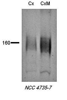
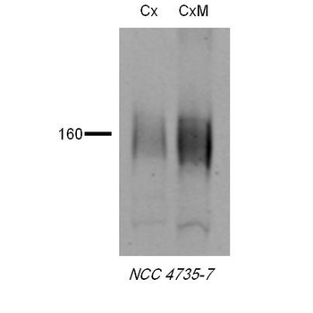
Western blot analysis of rat tissues using NCC antibody
Immunohistochemistry analysis using Rabbit Anti-NCC Polyclonal Antibody. Tissue: kidney tissue. Species: Rat. Primary Antibody: Rabbit Anti-NCC Polyclonal Antibody at 1:200. Secondary Antibody: FITC Goat Anti-Rabbit (green).
Western blot analysis of Rat tissue lysates showing detection of NCC protein using Rabbit Anti-NCC Polyclonal Antibody. Primary Antibody: Rabbit Anti-NCC Polyclonal Antibody at 1:1000.
Immunohistochemistry analysis using Rabbit Anti-NCC Polyclonal Antibody. Tissue: kidney tissue. Species: Mouse. Fixation: paraformaldehyde-fixed paraffin-embedded. Primary Antibody: Rabbit Anti-NCC Polyclonal Antibody at 1:500 for Overnight at 4C. Secondary Antibody: Biotinylated Goat Anti-Rabbit IgG at 1:500 for 30 min at RT.
Immunohistochemistry analysis using Rabbit Anti-NCC Polyclonal Antibody. Tissue: kidney tissue. Species: Mouse. Fixation: paraformaldehyde-fixed paraffin-embedded. Primary Antibody: Rabbit Anti-NCC Polyclonal Antibody at 1:1000 for Overnight at 4C. Secondary Antibody: Biotinylated Goat Anti-Rabbit IgG at 1:501 for 30 min at RT.
* VAT and and shipping costs not included. Errors and price changes excepted