Application Notes: 1 µg/ml was sufficient for detection of beta-ENaC in 20 µg of rat kidney tissue lysate by colorimetric immunoblot analysis using Goat anti-rabbit IgG:HRP as the secondary antibody
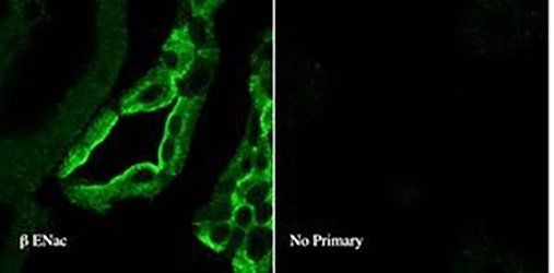
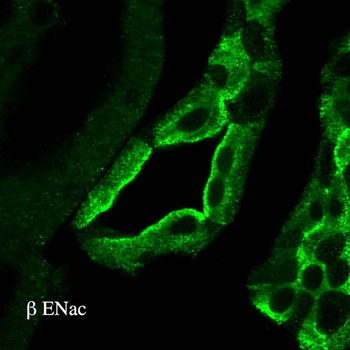
Confocal Immunofluorescencence analysis of rat kidney tissues using ENaC Beta antibody
Western blot analysis of rat kidney tissues using ENaC Beta antibody
Immunohistochemistry analysis using Rabbit Anti-ENaC Polyclonal Antibody. Tissue: kidney tissue. Species: Rat. Primary Antibody: Rabbit Anti-ENaC Polyclonal Antibody at 1:100. Secondary Antibody: FITC Goat Anti-Rabbit (green).
Western blot analysis of Rat kidney tissue lysates showing detection of ENaC protein using Rabbit Anti-ENaC Polyclonal Antibody. Primary Antibody: Rabbit Anti-ENaC Polyclonal Antibody at 1:1000.
Western blot analysis of Mouse mpkCCD cell lysates showing detection of ENaC protein using Rabbit Anti-ENaC Polyclonal Antibody. Primary Antibody: Rabbit Anti-ENaC Polyclonal Antibody at 1:1000.
Western blot analysis of Mouse kidney cortex showing detection of ENaC protein using Rabbit Anti-ENaC Polyclonal Antibody. Primary Antibody: Rabbit Anti-ENaC Polyclonal Antibody at 1:1000. Low-salt diet (lanes 1-4) compared to a high-salt diet (lanes 5-8).
* VAT and and shipping costs not included. Errors and price changes excepted