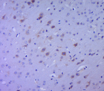
Paraformaldehyde-fixed, paraffin embedded (mouse brain tissue), Antigen retrieval by boiling in sodium citrate buffer (pH6.0) for 15 min, Block endogenous peroxidase by 3% hydrogen peroxide for 20 minutes, Blocking buffer (normal goat serum) at 37C for 30 min, Antibody incubation with (SPHK2) Polyclonal Antibody, Unconjugated (orb7003) at 1:400 overnight at 4C, followed by a conjugated secondary for 20 minutes and DAB staining.
Sample: Lane 1: Human SH-SY5Y cell Lysates, Lane 2: Human A549 cell Lysates, Lane 3: Human HepG2 cell Lysates, Primary: Anti-SPHK2 (orb7003) at 1/1000 dilution, Secondary: IRDye800CW Goat Anti-Rabbit IgG at 1/20000 dilution, Predicted band size: 72kDa, Observed band size: 72kDa.
Sample: Liver (Mouse) Lysate at 40 ug, Primary: Anti-SPHK2 (orb7003) at 1/300 dilution, Secondary: IRDye800CW Goat Anti-Rabbit IgG at 1/20000 dilution, Predicted band size: 72kD, Observed band size: 75kD.
Sample: Stomach (Mouse) Lysate at 40 ug, Primary: Anti-SPHK2 (orb7003) at 1/300 dilution, Secondary: IRDye800CW Goat Anti-Rabbit IgG at 1/20000 dilution, Predicted band size: 72kD, Observed band size: 75kD.
SH-SY5Y cell, 4% Paraformaldehyde-fixed, Triton X-100 at room temperature for 20 min, Blocking buffer (normal goat serum) at 37C for 20 min, Antibody incubation with (SPHK2) polyclonal Antibody, Unconjugated (orb7003) 1:100, 90 minutes at 37C, followed by a conjugated Goat Anti-Rabbit IgG antibody at 37C for 90 minutes, DAPI (blue) was used to stain the cell nuclei.
IHC-P analysis of mouse brain tissue using SPHK2 Polyclonal Antibody at 1:400 dilution.
* VAT and and shipping costs not included. Errors and price changes excepted