The subcellular fraction of human alveolar macrophages was used as the immunogen for this CD68 antibody.
Conjugation:
Unconjugated
This antibody recognizes a glycoprotein of 110kDa, which is identified as CD68. antibody to CD68 is important for identifying macrophages in tissue sections. It stains macrophages in a wide variety of human tissues, including Kupffer cells and macrophages in the red pulp of the spleen, in lamina propria of the gut, in lung alveoli, and in bone marrow. CD68 antibody reacts with myeloid precursors and peripheral blood granulocytes. It also reacts with plasmacytoid T cells, which are supposed to be of monocyte/macrophage origin. CD68 shows strong granular cytoplasmic staining of chronic and acute myeloid leukemia and also reacts with rare cases of true histiocytic neoplasia. Lymphomas are negative or show few granules.
Clonality:
Monoclonal
Clone Designation:
[C68/684]
Buffer:
0.2 mg/ml in 1X PBS with 0.1 mg/ml rAlbumin and 0.05% sodium azide
Application Dilute:
Western blot: 1-2ug/ml,Immunofluorescence: 1-2ug/ml,Flow cytometry: 1-2ug/million cells,Immunohistochemistry (FFPE): 0.5-1ug/ml for 30 min at RT
Application Notes:
Application Notes: The concentration stated for each application is a general starting point. Variations in protocols, secondaries and substrates may require the antibody to be titered up or down for optimal performance.1. Staining of formalin-fixed tissues is enhanced by boiling tissue sections in 10mM Citrate Buffer, pH 6 or pH 9 10mM Tris with 1mM EDTA for 10-20 min followed by cooling at RT for 20 minutes.2. The prediluted format is supplied in a dropper bottle and is optimized for use in IHC. After epitope retrieval step (if required), drip mAb solution onto the tissue section and incubate at RT for 30 min
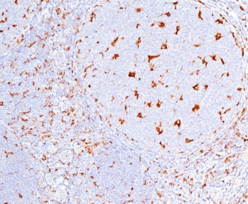
IHC testing of human tonsil (10X) stained with CD68 antibody (C68/684).
Formalin-fixed, paraffin-embedded human Histiocytoma stained with CD68 antibody.
Immunofluorescent staining of fixed human U-87 MG cells with CD68 antibody (clone C68/684, green) and Reddot nuclear stain (red).
Flow cytometry testing of fixed human U-87 MG cells with CD63 antibody (clone C68/684), Red = isotype control, Blue = CD63 antibody.
Western blot testing of human spleen lysate with CD68 antibody (clone C68/684). Expected molecular weight: 37-110 kDa depending on glycosylation level.
SDS-PAGE analysis of purified, BSA-free CD68 antibody (clone C68/684) as confirmation of integrity and purity.
* VAT and and shipping costs not included. Errors and price changes excepted