CD31, also called PECAM-1, is a transmembrane glycoprotein member of the immunoglobulin supergene family of adhesion molecules. It is expressed by stem cells of the hematopoietic system and CD31 antibody can be used to identify and concentrate these cells for experimental studies as well as for bone marrow transplantation. CD31 antibody has shown to be highly specific and sensitive for vascular endothelial cells. Staining of nonvascular tumors (excluding hematopoietic neoplasms) is rare. This antibody reacts with normal, benign, and malignant endothelial cells which make up blood vessel lining. The level of CD31 expression can help to determine the degree of tumor angiogenesis, and a high expression level may imply a rapidly growing tumor and potentially a predictor of tumor recurrence.
Clonality:
Monoclonal
Clone Designation:
[C31.3]
Buffer:
0.2 mg/ml in 1X PBS with 0.1 mg/ml rAlbumin and 0.05% sodium azide
Application Dilute:
Flow cytometry: 1-2ug/10 6 cells,Western blot: 1-2ug/ml,Immunofluorescence: 1-2ug/ml,Immunohistochemistry (FFPE): 1-2ug/ml for 30 min at RT
Application Notes:
Application Notes: Differences in protocols, secondaries and substrates may require the CD31 antibody to be titered for optimal performance.1. Staining of formalin-fixed tissues requires boiling tissue sections in 1mM EDTA, pH 7.5-8.5, for 10-20 min followed by cooling at RT for 20 minutes.2. The prediluted format is supplied in a dropper bottle and is optimized for use in IHC. After epitope retrieval step (if required), drip mAb solution onto the tissue section and incubate at RT for 30 min
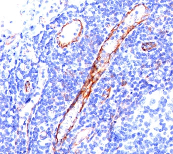
IHC staining of tonsil tissue with CD31 antibody (C31.3).
IHC staining of angiocarcinoma with CD31 antibody (C31.3).
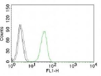
FACS testing of Jurkat cells with Alexa Fluor conjugated CD31 antibody (green) and isotype control (gray).
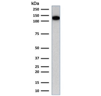
Western blot testing of human Jurkat cell lysate with CD31 antibody (clone C31.1). Expected molecular weight: 83-130 kDa depending on level of glycosylation.
Immunofluorescent staining of PFA-fixed human Jurkat cells with CD31 antibody (green, clone C31.3) and NucSpot nuclear counterstain (red).
Western blot testing of human ThP-1 cell lysate with CD31 antibody (clone C31.1). Expected molecular weight: 83-130 kDa depending on level of glycosylation.
* VAT and and shipping costs not included. Errors and price changes excepted