Recombinant human HSPB1 protein was used as the immunogen for the HSP27 antibody.
Conjugation:
Unconjugated
It recognizes a 24-27kDa estrogen-regulated protein, identified as heat shock protein 27 (hsp27). Hsp27 was recently found to be identical to the estrogen-induced p29 and 24K protein. About 50% of breast carcinomas are positive for hsp27 especially those that are also positive for estrogen and/or progesterone receptor. HSP27 has also been implicated in drug resistance in cancer cells.
0.2 mg/ml in 1X PBS with 0.1 mg/ml rAlbumin and 0.05% sodium azide
Application Dilute:
Flow cytometry: 1-2ug/million cells,Immunofluorescence: 1-2ug/ml,Immunohistochemistry (FFPE): 1-2ug/ml for 30 min at RT,Western blot: 1-2ug/ml
Application Notes:
Application Notes: Optimal dilution of the HSP27 antibody should be determined by the researcher.1. Staining of formalin-fixed tissues requires boiling tissue sections in pH 9 10mM Tris with 1mM EDTA for 10-20 min followed by cooling at RT for 20 min2. The prediluted format is supplied in a dropper bottle and is optimized for use in IHC. After epitope retrieval step (if required), drip mAb solution onto the tissue section and incubate at RT for 30 min
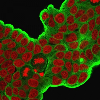
Immunofluorescent staining of PFA-fixed human MCF7 cells with HSP27 antibody (clone HSPB1/774, green) and Reddot nuclear stain (red).
IHC: Formalin-fixed, paraffin-embedded human breast carcinoma stained with HSP27 antibody (clone HSPB1/774).
IHC: Formalin-fixed, paraffin-embedded human prostate carcinoma stained with HSP27 antibody (clone HSPB1/774).
Flow cytometry testing of PFA-fixed human MCF7 cells with HSP27 antibody (clone HSPB1/774), Red = isotype control, Blue = HSP27 antibody.
SDS-PAGE analysis of purified, BSA-free HSP27 antibody (clone HSPB1/774) as confirmation of integrity and purity.
Western blot testing of human HeLa cell lysate with HSP27 antibody. Expected molecular weight: 23~27 kDa.
* VAT and and shipping costs not included. Errors and price changes excepted