
TOP1 Recombinant Monoclonal Antibody, Clone: [6D8], Unconjugated, Rabbit
Catalog Number:
CSB-RA792129A0HU
- Images (5)
| Article Name: | TOP1 Recombinant Monoclonal Antibody, Clone: [6D8], Unconjugated, Rabbit |
| Biozol Catalog Number: | CSB-RA792129A0HU |
| Supplier Catalog Number: | CSB-RA792129A0HU |
| Alternative Catalog Number: | CSB-RA792129A0HU-100UL, CSB-RA792129A0HU-50UL |
| Manufacturer: | Cusabio |
| Host: | Rabbit |
| Category: | Antikörper |
| Application: | ELISA, FC, IHC, IP, WB |
| Species Reactivity: | Human |
| Conjugation: | Unconjugated |
| Alternative Names: | DNA topoisomerase 1 (EC 5.99.1.2) (DNA topoisomerase I) , TOP1 |
| Clonality: | Monoclonal |
| Clone Designation: | [6D8] |
| UniProt: | P11387 |
| Buffer: | Rabbit IgG in 10mM phosphate buffered saline , pH 7.4, 150mM sodium chloride, 0.05% BSA, 0.02% sodium azide and 50% glycerol. |
| Purity: | Affinity-chromatography |
| Form: | Liquid |
| Target: | TOP1 |
| Antibody Type: | Recombinant Antibody |
| Application Dilute: | Recommended dilution: WB:1:500-1:5000, IHC:1:50-1:200, FC:1:20-1:200, IP:1:200-1:1000 |
|
|
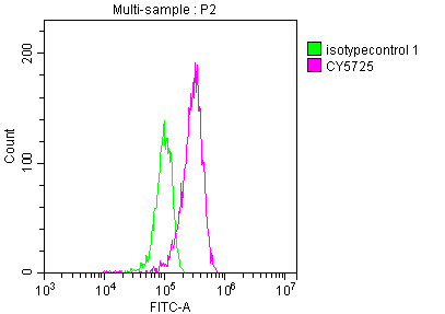
Overlay histogram showing HepG2 cells stained with CSB-RA792129A0HU (red line) at 1:50. The cells were fixed with 70% Ethylalcohol (18h) and then incubated in 10% normal goat serum to block non-specific protein-protein interactions followedby the antibody (1µg/1*106 cells) for 1 h at 4°C.The secondary antibody used was FITC-conjugated goat anti-rabbit IgG (H+L) at 1/200 dilution for 30min at 4°C. Control antibody (green line) was Rabbit IgG (1µg/1*106 cells) used under the same conditions. Acquisition of >10,000 events was performed. |
|
|
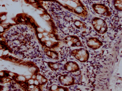
IHC image of CSB-RA792129A0HU diluted at 1:100 and staining in paraffin-embedded human small intestine tissue performed on a Leica BondTM system. After dewaxing and hydration, antigen retrieval was mediated by high pressure in a citrate buffer (pH 6.0) . Section was blocked with 10% normal goat serum 30min at RT. Then primary antibody (1% BSA) was incubated at 4°C overnight. The primary is detected by a Goat anti-rabbit IgG polymer labeled by HRP and visualized using 0.05% DAB. |
|
|
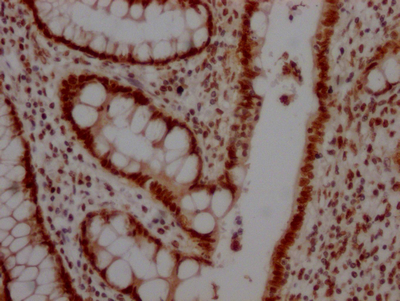
IHC image of CSB-RA792129A0HU diluted at 1:100 and staining in paraffin-embedded human colon cancer performed on a Leica BondTM system. After dewaxing and hydration, antigen retrieval was mediated by high pressure in a citrate buffer (pH 6.0) . Section was blocked with 10% normal goat serum 30min at RT. Then primary antibody (1% BSA) was incubated at 4°C overnight. The primary is detected by a Goat anti-rabbit IgG polymer labeled by HRP and visualized using 0.05% DAB. |
|
|
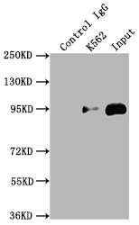
Immunoprecipitating TOP1 in K562 whole cell lysate Lane 1: Rabbit control IgG instead of CSB-RA792129A0HU in K562 whole cell lysate.For western blotting,a HRP-conjugated Protein G antibody was used as the secondary antibody (1/2000) Lane 2: CSB-RA792129A0HU(2µg) + K562 whole cell lysate(500µg) Lane 3: K562 whole cell lysate (10µg) |
|
|
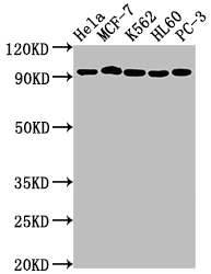
Western Blot Positive WB detected in: Hela whole cell lysate, MCF-7 whole cell lysate, K562 whole cell lysate, HL60 whole cell lysate, PC-3 whole cell lysate All lanes: TOP1 antibody at 1:2000 Secondary Goat polyclonal to rabbit IgG at 1/50000 dilution Predicted band size: 91 kDa Observed band size: 91 kDa |
Product Guarantee and Expert Support
