
CLSTN1 Recombinant Monoclonal Antibody, Clone: [11H4], Unconjugated, Rabbit
Catalog Number:
CSB-RA923470A0HU
- Images (5)
| Article Name: | CLSTN1 Recombinant Monoclonal Antibody, Clone: [11H4], Unconjugated, Rabbit |
| Biozol Catalog Number: | CSB-RA923470A0HU |
| Supplier Catalog Number: | CSB-RA923470A0HU |
| Alternative Catalog Number: | CSB-RA923470A0HU-100UL, CSB-RA923470A0HU-50UL |
| Manufacturer: | Cusabio |
| Host: | Rabbit |
| Category: | Antikörper |
| Application: | ELISA, FC, IF, IHC, WB |
| Species Reactivity: | Human, Mouse, Rat |
| Conjugation: | Unconjugated |
| Alternative Names: | Alc alpha antibody, Alc-alpha antibody, Alcadein alpha 1 antibody, Alcadein alpha antibody, Alcadein-alpha antibody, alcalpha1 antibody, alcalpha2 antibody, Alzheimer related cadherin like protein antibody, Alzheimer-related cadherin-like protein antibody, C-terminal fragment 1-alpha antibody, Cadherin related family member 12 antibody, Calsyntenin 1 antibody, Calsyntenin1 antibody, CDHR12 antibody, CLSTN 1 antibody, Clstn1 antibody, CS1 antibody, CSTN1 antibody, CSTN1_HUMAN antibody, CTF1-alpha antibody, FLJ32258 antibody, KIAA0911 antibody, Non classical cadherin XB31alpha antibody, Non classical cadherin XB31alpha1 antibody, Non-classical cadherin XB31alpha antibody, PIK3CD antibody, SAlc-alpha antibody, XB31alpha antibody |
| Clonality: | Monoclonal |
| Clone Designation: | [11H4] |
| UniProt: | O94985 |
| Buffer: | Rabbit IgG in 10mM phosphate buffered saline , pH 7.4, 150mM sodium chloride, 0.05% BSA, 0.02% sodium azide and 50% glycerol. |
| Purity: | Affinity-chromatography |
| Form: | Liquid |
| Target: | CLSTN1 |
| Antibody Type: | Recombinant Antibody |
| Application Dilute: | Recommended dilution: WB:1:500-1:5000, IHC:1:50-1:200, IF:1:50-1:200, FC:1:50-1:200 |
|
|
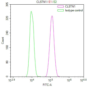
Overlay Peak curve showing Hela cells stained with CSB-RA923470A0HU (red line) at 1:100. The cells were fixed in 4% formaldehyde and permeated by 0.2% TritonX-100 for10min. Then 10% normal goat serum to block non-specific protein-protein interactions followed by the antibody (1ug/1*106cells) for 45min at 4°C. The secondary antibody used was FITC-conjugated goat anti-rabbit IgG (H+L) at 1/200 dilution for 35min at 4°C.Control antibody (green line) was Rabbit IgG (1ug/1*106cells) used under the same conditions. Acquisition of >10, 000 events was performed. |
|
|
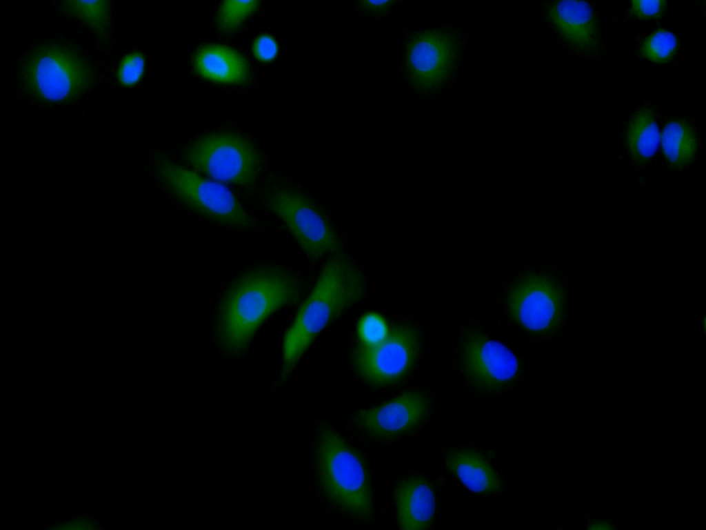
Immunofluorescence staining of A549 cell with CSB-RA923470A0HU at 1:50 , counter-stained with DAPI. The cells were fixed in 4% formaldehyde, permeabilized using 0.2% Triton X-100 and blocked in 10% normal Goat Serum. The cells were then incubated with the antibody overnight at 4C. The secondary antibody was Alexa Fluor 488-congugated AffiniPure Goat Anti-Rabbit IgG(H+L). |
|
|
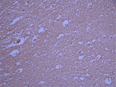
IHC image of CSB-RA923470A0HU diluted at 1:100 and staining in paraffin-embedded human brain tissue performed on a Leica BondTM system. After dewaxing and hydration, antigen retrieval was mediated by high pressure in a citrate buffer (pH 6.0). Section was blocked with 10% normal goat serum 30min at RT. Then primary antibody (1% BSA) was incubated at 4C overnight. The primary is detected by a Goat anti-rabbit polymer IgG labeled by HRP and visualized using 0.05% DAB. |
|
|
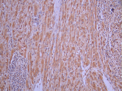
IHC image of CSB-RA923470A0HU diluted at 1:100 and staining in paraffin-embedded human liver cancer performed on a Leica BondTM system. After dewaxing and hydration, antigen retrieval was mediated by high pressure in a citrate buffer (pH 6.0). Section was blocked with 10% normal goat serum 30min at RT. Then primary antibody (1% BSA) was incubated at 4C overnight. The primary is detected by a Goat anti-rabbit polymer IgG labeled by HRP and visualized using 0.05% DAB. |
|
|
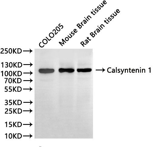
Western Blot Positive WB detected in: COLO205 whole cell lysate(30µg), Mouse brain tissue lysate(30µg), Rat brain tissue lysate(30µg) All lanes:Calsyntenin 1 antibody at 1:1000 Secondary Goat polyclonal to rabbit IgG at 1/40000 dilution Predicted band size: 110 kDa Observed band size: 110 kDa Exposure time:1min |
Product Guarantee and Expert Support
