The antiserum was produced against synthesized peptide derived from human DIL-2. AA range:301-350
Alternative Names:
TPX2, C20orf1, C20orf2, DIL2, HCA519, Targeting protein for Xklp2, Differentially expressed in cancerous and non-cancerous lung cells 2, DIL-2, Hepatocellular carcinoma-associated antigen 519, Protein fls353, Restricted expression prolifera
developmental stage:Exclusively expressed in proliferating cells from the transition G1/S until the end of cytokinesis.,PTM:Phosphorylated upon DNA damage, probably by ATM or ATR.,subcellular location:During mitosis it is strictly associated with the spindle pole and with the mitotic spindle, whereas during S and G2, it is diffusely distributed throughout the nucleus.,tissue specificity:Expressed in lung carcinoma cell lines but not in normal lung tissues.,
Western Blot: 1/500 - 1/2000. Immunohistochemistry: 1/100 - 1/300. ELISA: 1/10000. Not yet tested in other applications.
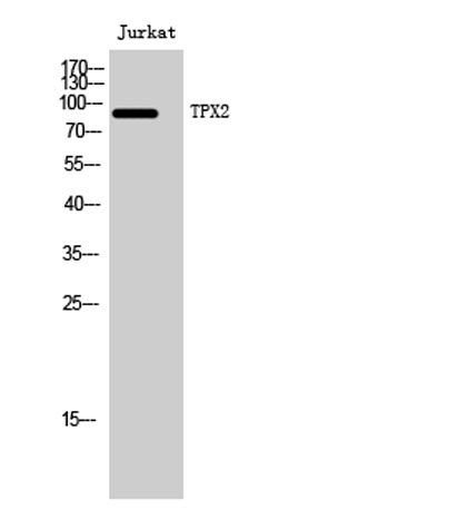
Western Blot analysis of Jurkat cells using TPX2 Polyclonal Antibody. Secondary antibody(catalog:RS0002) was diluted at 1:20000 cells nucleus extracted by Minute TM Cytoplasmic and Nuclear Fractionation kit (SC-003,Inventbiotech,MN,USA).
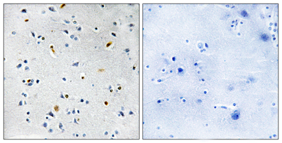
Immunohistochemistry analysis of paraffin-embedded human brain tissue, using DIL-2 Antibody. The picture on the right is blocked with the synthesized peptide.
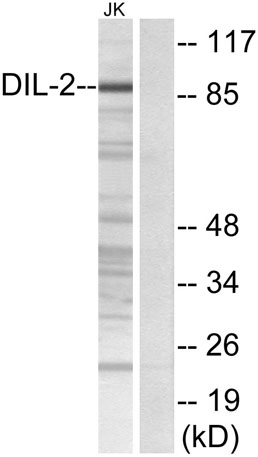
Western blot analysis of lysates from Jurkat cells, using DIL-2 Antibody. The lane on the right is blocked with the synthesized peptide.
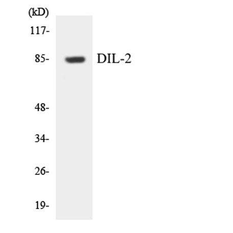
Western blot analysis of the lysates from HUVECcells using DIL-2 antibody.
* VAT and and shipping costs not included. Errors and price changes excepted