Anti-Rabbit IgG FITC, TrueBlot, FITC TrueBlot ULTRA, Fluorescein TrueBlot, TrueBlot for IP/WB, TrueBlot for immunoprecipitation, TrueBlot for western blotting, Fluorescent TrueBlot, Rb TrueBlot
Clonality:
Monoclonal
Concentration:
1.0 mg/mL by UV absorbance at 280 nm
Clone Designation:
[eB182]
Buffer:
0.02 M Potassium Phosphate, 0.15 M Sodium Chloride, pH 7.2
Form:
Lyophilized
Target:
Rabbit
Application Dilute:
FLISA: User Optimized, Flow Cytometry: 1:2,000 - 1:10,000, IHC: User Optimized, IF Microscopy: 1:500 - 1:2,500, WB: 1:1000
Application Notes:
Rabbit IgG TrueBlot Fluorescein Conjugated Antibody has been tested in immunofluorescence microscopy, fluorescent western blotting, and immunoprecipitation and are suitable for fluorescence based plate assays (FLISA, multiplex analysis, including multic
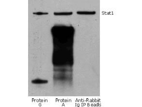
Rabbit TrueBlot IP / Western Blot: Jurkat cell lysate (0.5 ml of 1x10e7 cells/ml) was incubated with rabbit anti-human Stat1 and immunoprecipitated using Protein G, Protein A and Anti-Rabbit Ig IP Beads. Precipitate from 5x10e5 cells was subjected to electrophoresis, transferred to a PVDF membrane, and Western blotted with anti-Stat1 using Rabbit TrueBlot: Anti-Rabbit IgG HRP
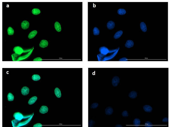
Immunofluorescence microscopy of BCL3 in Caco-2 cells using FITC-conjugated Fluorescent TrueBlot anti-rabbit IgG for detection. Caco-2 cells were fixed with 4% PFA, blocked (5% mouse serum/0.3% Triton X-100 in 1X PBS ) for 1 hr, then incubated with 15 µg/mL of anti-BCL3 primary antibody (Cat. No. 600-401-GU4) at 4C overnight. Following 3 washes in 1X PBS for 5 min each, 5 µg/mL of FITC-conjugated Fluorescent TrueBlot anti-rabbit IgG was added and allowed to incubate for 1 hr at room temperature. Nuclei were counterstained with DAPI present in mounting medium. The predicted main localization is nucleoplasm. Additional localization in some cell types includes vesicles and midbody. (a) BCL3 (b) DAPI (c) merged DAPI/BCL3 (d) secondary antibody only. Image taken at 40X magnification.
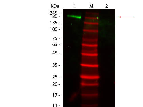
Western Blot of Fluorescent TrueBlot: Anti-Rabbit IgG Fluorescein. Lane 1: Rabbit IgG, Non-reduced. Lane 2: Rabbit IgG, Reduced. Load: 50 ng per lane. Primary antibody: none. Secondary antibody: Fluorescent TrueBlot: Anti-Rabbit IgG Fluorescein at 1:1,000 for 60 min at RT. Block: MB-070 for 30 min at RT. Predicted/Observed size: 160 kDa for Rabbit IgG, Non-reduced. Other band(s): none.
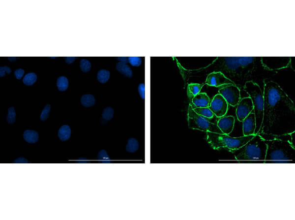
Immunofluorescence microscopy of ZO-1 in Caco-2 cells using FITC-conjugated Fluorescent TrueBlot anti-rabbit IgG for detection. Caco-2 cells were fixed with 4% PFA, blocked (5% mouse serum/0.3% Triton X-100 in 1X PBS) for 1 hr, then incubated with 15 µg/mL of anti-ZO-1 primary antibody (Cat. No. 600-401-GU7) at 4C overnight. Following 3 washes in 1X PBS for 5 min each, 5 µg/mL of FITC-conjugated Fluorescent TrueBlot anti-rabbit IgG was added and allowed to incubate for 1 hr at room temperature. Nuclei were counterstained with DAPI present in mounting medium. Predicted cell localization is cell membrane and cell junctions. Image taken at 40X magnification. (right) Merged DAPI (blue)/ZO-1 (green), image shown (left) secondary antibody only.
* VAT and and shipping costs not included. Errors and price changes excepted